A guide to selecting high-performing antibodies for S1PR1 (UniProt ID: P21453) for use in western blot, immunoprecipitation, and immunofluorescence
Riham Ayoubi, Maryam Fotouhi, Charles Alende, Sara González Bolívar, Kathleen Southern, Carl Laflamme, Melissa Pitman, Kathleen Southern

TL;DR
This paper evaluates nine commercial antibodies for S1PR1 to help researchers choose the best ones for common lab techniques.
Contribution
The study provides a standardized evaluation of S1PR1 antibodies to improve reproducibility in research.
Findings
Nine S1PR1 antibodies were tested for western blot, immunoprecipitation, and immunofluorescence.
Results are shared openly to help researchers select reliable antibodies for their experiments.
Abstract
Sphingosine 1-phosphate receptor 1 (S1PR1) is a G-coupled protein receptor that induces crucial biological processes when bound by sphingosine 1-phosphate. Here, we have characterized nine S1PR1 commercial antibodies for western blot, immunoprecipitation, and immunofluorescence using a standardized experimental protocol based on comparing read-outs in knockout cell lines and isogenic parental controls. These studies are part of a larger, collaborative initiative seeking to address antibody reproducibility issues by characterizing commercially available antibodies for human proteins and publishing the results openly as a resource for the scientific community. While use of antibodies and protocols vary between laboratories, we encourage readers to use this report as a guide to select the most appropriate antibodies for their specific needs.
Genes, proteins, chemicals, diseases, species, mutations and cell lines named across the full text — each resolved to its canonical identifier and authoritative record.
Click any figure to enlarge with its caption.
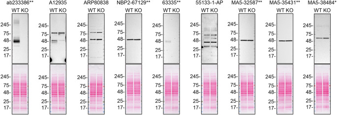 Figure 1
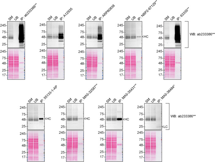 Figure 2
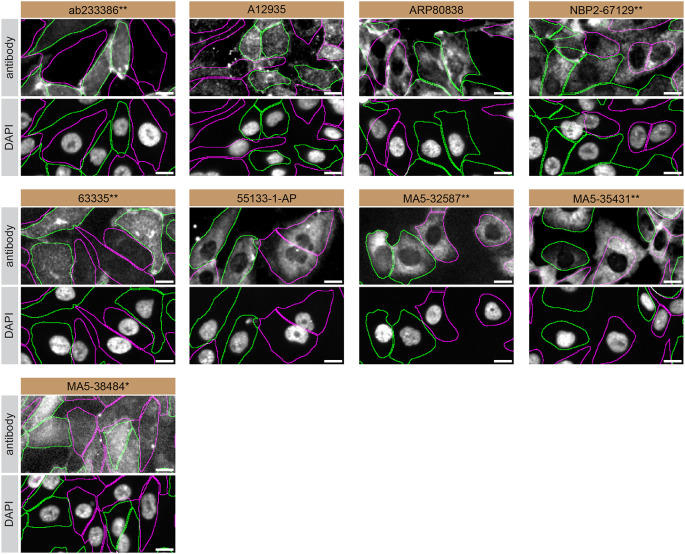 Figure 3
| Institution | Catalog number | RRID (Cellosaurus) | Cell line | Genotype |
|---|---|---|---|---|
| ATCC | HTB-52 |
| SK-HEP-1 | WT |
| Abcam | - | - | SK-HEP-1 |
|
| Company | Catalog number | Lot number | RRID (Antibody Registry) | Clonality | Clone ID | Host | Concentration (μg/μl) | Vendors recommended applications |
|---|---|---|---|---|---|---|---|---|
| Abcam | ab233386
| GR3404607-2 |
| recombinant-mono | rabbit | 0.43 | Wb | |
| ABclonal | A12935 | 59370101 |
| polyclonal | - | rabbit | 1.10 | Wb |
| Aviva Systems Biology | QC56393-190325 |
| polyclonal | - | rabbit | 0.50 | Wb | |
| Novus Biologicals (a Bio-Techne brand) | NBP2-67129
| HN1019 |
| recombinant-mono | JM10-66 | rabbit | 1.00 | Wb, IF |
| Cell Signaling Technology | 63335
| 1 |
| recombinant-mono | E8U3O | rabbit | 0.20 | Wb, IP, IF |
| Proteintech | 55133-1-AP | 89828 |
| polyclonal | - | rabbit | 1.00 | Wb, IP |
| Thermo Fisher Scientific | MA5-32587
| XC3523726 |
| recombinant-mono | JM10-66 | rabbit | 1.00 | Wb, IF |
| Thermo Fisher Scientific | MA5-35431
| XC3523881 |
| recombinant-mono | ARC0881 | rabbit | 0.29 | Wb |
| Thermo Fisher Scientific | MA5-38484
| XC3523353 |
| monoclonal | 8EAH5 | mouse | 1.00 | Wb |
- —Ontario Genomics
- —Genome Quebec
- —Emory-Sage-SGC TaRget Enablement to Accelerate Therapy Development for Alzheimer’s Disease
- —National Institute on Aging
- —Mitacs
- —Genome Canada
Peer Reviews
No public reviews on file for this paper yet. If you reviewed it on a platform where reviews are public (OpenReview, ICLR, NeurIPS, ICML), you can paste yours below so the community can read it here.
Videos
No videos yet. Explain this paper in a talk, walkthrough, or lecture? Add one.
Taxonomy
TopicsSphingolipid Metabolism and Signaling · Cellular transport and secretion · Erythrocyte Function and Pathophysiology
Introduction
Sphingosine 1-phosphate receptor 1 (S1PR1), is a G-protein coupled receptor which binds to its abundant ligand, sphingosine 1-phosphate, inducing intracellular signalling pathways in cell growth, differentiation, migration and trafficking. ^ 1 ^ ^–^ ^ 4 ^ S1PR1 activation by sphingosine 1-phosphate is essential for neuronal events, and its dysregulation may contribute to the pathogenesis of Alzheimer’s disease. ^ 5 ^ ^,^ ^ 6 ^ High-performing S1PR1 antibodies would facilitate S1PR1 research and uncover therapeutic strategies.
This research is part of a broader collaborative initiative in which academics, funders and commercial antibody manufacturers are working together to address antibody reproducibility issues by characterizing commercial antibodies for human proteins using standardized protocols, and openly sharing the data. ^ 7 ^ ^–^ ^ 9 ^ Here we evaluated the performance of nine commercial antibodies for S1PR1 for use in western blot, immunoprecipitation, and immunofluorescence, enabling biochemical and cellular assessment of S1PR1 properties and function. The platform for antibody characterization used to carry out this study was endorsed by a committee of industry and academic representatives. It involves identifying appropriate cell lines with adequate target protein expression, developing or contributing equivalent knockout (KO) cell lines and finally, characterizing most commercially available antibodies against the corresponding target protein. The standardized antibody characterization protocols are openly available on Protocol Exchange (DOI: 10.21203/rs.3.pex-2607/v1). ^ 10 ^
The authors do not engage in result analysis or offer explicit antibody recommendations. A limitation of this study is the use of universal protocols - any conclusions remain relevant within the confines of the experimental setup and cell line used in this study. Our primary aim is to deliver top-tier data to the scientific community, grounded in Open Science principles. This empowers experts to interpret the characterization data independently, enabling them to make informed choices regarding the most suitable antibodies for their specific experimental needs. Guidelines on how to interpret antibody characterization data found in this study are featured on the YCharOS gateway. ^ 11 ^
Results and discussion
Our standard protocol involves comparing readouts from WT (wild type) and KO cells. ^ 12 ^ ^,^ ^ 13 ^ The first step is to identify a cell line(s) that expresses sufficient levels of S1PR1 to generate a measurable signal using antibodies. To this end, we examined the DepMap transcriptomics database to identify all cell lines that express the target at levels greater than 2.5 log 2 (transcripts per million “TPM” + 1), which we have found to be a suitable cut-off (Cancer Dependency Map Portal, RRID:SCR_017655). The SK-HEP-1 cells expresses the S1PR1 transcript at 6.5 log 2 (TPM+1) RNA levels, which is above the average range of cancer cells analyzed. Parental and S1PR1 KO SK-HEP-1 cells were obtained from Abcam ( Table 1).
For western blot experiments, WT and S1PR1 KO protein lysates were rain on SDS-PAGE, transferred onto nitrocellulose membranes, and then probed with nine S1PR1 antibodies in parallel ( Table 2, Figure 1).
S1PR1 antibody screening by western blot.Lysates of SK-HEP-1 (WT and S1PR1 KO) were prepared and 30 μg of protein were processed for western blot with the indicated S1PR1 antibodies. The Ponceau stained transfers of each blot are presented to show equal loading of WT and KO lysates and protein transfer efficiency from precast midi 4-20% Tris-Glycine polyacrylamide gel (Thermo Fisher Scientific, cat number WXP42012BOX) to the nitrocellulose membrane. Antibody dilutions were chosen according to the recommendations of the antibody supplier. Antibody dilution used: ab233386* at 1/1000, A12935 at 1/1000, ARP80838 at 1/500., NBP2-67129** at 1/1000, 63335** at 1/1000, 55133-1-AP at 1/1000, MA5-32587** at 1/1000, MA5-35431** at 1/1000, MA5-38484* at 1/500. Predicted band size: 42.8 kDa. *Monoclonal antibody, *Recombinant antibody.
We then assessed the capability of all nine antibodies to capture S1PR1 from SK-HEP-1 protein extracts using immunoprecipitation techniques, followed by western blot analysis. For the immunoblot step, a specific S1PR1 antibody identified previously (refer to Figure 1) was selected. Equal amounts of the starting material (SM), the unbound fraction (UB), as well as the whole immunoprecipitate (IP) eluates were separated by SDS-PAGE ( Figure 2).
S1PR1 antibody screening by immunoprecipitation.SK-HEP-1 lysates were prepared, and immunoprecipitation was performed using 2.0 μg of the indicated S1PR1 antibodies pre-coupled to Dynabeads protein A or protein G. Samples were washed and processed for western blot on a precast midi 4-20% Tris-Glycine polyacrylamide gel with the indicated S1PR1 antibodies. For western blot, ab233386* was used at 1/1000. The Ponceau stained transfers of each blot are shown. SM = 4% starting material; UB = 4% unbound fraction; IP = immunoprecipitate, HC = antibody heavy chain, LC = antibody light chain. *Monoclonal antibody, *Recombinant antibody.
For immunofluorescence, nine antibodies were screened using a mosaic strategy. First, SK-HEP-1 WT and S1PR1 KO cells were labelled with different fluorescent dyes in order to distinguish the two cell lines, and the S1PR1 antibodies were evaluated. Both WT and KO cells were imaged in the same field of view to reduce staining, imaging and image analysis bias ( Figure 3). Quantification of immunofluorescence intensity in hundreds of WT and KO cells was performed for each antibody tested, ^ 10 ^ and the images presented in Figure 3 are representative of this analysis.
S1PR1 antibody screening by immunofluorescence.SK-HEP-1 WT and S1PR1 KO cells were labelled with a green or a far-red fluorescent dye, respectively. WT and KO cells were mixed and plated to a 1:1 ratio in a 96-well plate with optically clear flat-bottom. Cells were stained with the indicated S1PR1 antibodies and with the corresponding Alexa-fluor 555 coupled secondary antibody including DAPI. Acquisition of the blue (nucleus-DAPI), green (identification of WT wells), red (antibody staining) and far-red (identification of KO cells) channels was performed. Representative images of the merged blue and red (grayscale) channels are shown. WT and KO cells are outlined with green and magenta dashed line, respectively. When an antibody was recommended for immunofluorescence by the supplier, we tested it at the recommended dilution. The rest of the antibodies were tested at 1 and 2 μg/ml and the final concentration was selected based on the detection range of the microscope used and a quantitative analysis not shown here. Antibody dilution used: ab233386* at 1/400, A12935 at 1/1000, ARP80838 at 1/250., NBP2-67129** at 1/1000, 63335** at 1/2000, 55133-1-AP at 1/500, MA5-32587** at 1/1000, MA5-35431** at 1/300, MA5-38484* at 1/200. Bars = 10 μm. *Monoclonal antibody, *Recombinant antibody.
In conclusion, we have screened nine S1PR1 commercial antibodies by western blot, immunoprecipitation, and immunofluorescence by comparing the signal produced by the antibodies in human SK-HEP-1 WT and S1PR1 KO cells. Several high-quality antibodies that successfully detect S1PR1 under our standardized experimental protocol can be identified. Researchers who wish to study S1PR1 in a different species are encouraged to select high-quality antibodies, based on the results of this study, and investigate the predicted species reactivity of the manufacturer before extending their research.
The underlying data for this study can be found on Zenodo, an open-access repository for which YCharOS has its own collection of antibody characterization reports. ^ 14 ^ ^,^ ^ 15 ^
Methods
The standardized protocols used to carry out this KO cell line-based antibody characterization platform was established and approved by a collaborative group of academics, industry researchers and antibody manufacturers. The detailed materials and step-by-step protocols used to characterize antibodies in western blot, immunoprecipitation and immunofluorescence are openly available on Protocol Exchange, a preprint server (DOI: 10.21203/rs.3.pex-2607/v1). ^ 10 ^
Antibodies and cell line used
Cell lines used and primary antibodies tested in this study are listed in Tables 1 and 2, respectively. To ensure that the cell lines and antibodies are cited properly and can be easily identified, we have included their corresponding Research Resource Identifiers, or RRID. ^ 16 ^ ^,^ ^ 17 ^
The reference list from the paper itself. Each links out to its DOI / PubMed record.
- 1Hla T Maciag T : An abundant transcript induced in differentiating human endothelial cells encodes a polypeptide with structural similarities to G-protein-coupled receptors. J. Biol. Chem. 1990;265(16):9308–9313. 10.1016/S 0021-9258(19)38849-0 2160972 · doi ↗ · pubmed ↗
- 2Lee MJ Van Brocklyn JR Thangada S : Sphingosine-1-phosphate as a ligand for the G protein-coupled receptor EDG-1. Science. 1998;279(5356):1552–1555. 10.1126/science.279.5356.1552 9488656 · doi ↗ · pubmed ↗
- 3Bryan AM Del Poeta M : Sphingosine-1-phosphate receptors and innate immunity. Cell. Microbiol. 2018;20(5):e 12836. 10.1111/cmi.12836 29498184 PMC 5893408 · doi ↗ · pubmed ↗
- 4Pyne S Pyne NJ : Translational aspects of sphingosine 1-phosphate biology. Trends Mol. Med. 2011;17(8):463–472. 10.1016/j.molmed.2011.03.002 21514226 · doi ↗ · pubmed ↗
- 5Jung Y Lopez-Benitez J Tognoni CM : Dysregulation of sphingosine-1-phosphate (S 1P) and S 1P receptor 1 signaling in the 5x FAD mouse model of Alzheimer’s disease. Brain Res. 2023;1799:148171. 10.1016/j.brainres.2022.148171 36410428 · doi ↗ · pubmed ↗
- 6Blaho VA Hla T : An update on the biology of sphingosine 1-phosphate receptors. J. Lipid Res. 2014;55(8):1596–1608. 10.1194/jlr.R 046300 24459205 PMC 4109755 · doi ↗ · pubmed ↗
- 7Ayoubi R Ryan J Biddle MS : Scaling of an antibody validation procedure enables quantification of antibody performance in major research applications. elife. 2023;12:12. 10.7554/e Life.91645.2 PMC 1066693137995198 · doi ↗ · pubmed ↗
- 8Carter AJ Kraemer O Zwick M : Target 2035: probing the human proteome. Drug Discov. Today. 2019;24(11):2111–2115.31278990 10.1016/j.drudis.2019.06.020 · doi ↗ · pubmed ↗