Multi-sequence MRI based radiomics nomogram for prediction expression of programmed death ligand 1 in thymic epithelial tumor
Jie Shen, Lantian Zhang, Shuke Li, Xiaofei Mu, Tongfu Yu, Wei Zhang, Yue Yu, Jing He, Wen Gao

TL;DR
This study develops a radiomics nomogram using MRI scans to predict PD-L1 expression in thymic epithelial tumors, offering a non-invasive alternative to traditional methods.
Contribution
A novel radiomics nomogram combining MRI features and clinical variables is proposed for predicting PD-L1 status in thymic epithelial tumors.
Findings
A radiomics signature with four features effectively differentiates PD-L1 positive and negative TET patients.
The combined radiomics nomogram achieved high AUC values (0.903 in training, 0.894 in validation) for PD-L1 prediction.
Calibration and decision curve analyses confirmed the clinical usefulness of the integrated model.
Abstract
High expression levels of programmed death receptor 1 (PD-1) and its ligand 1 (PD-L1) have been observed in thymic epithelial tumors (TET), suggesting their potential as prognostic indicators for disease progression and the effectiveness of immunotherapy in TET. The conventional method obtaining PD-L1 was challenging due to invasive sampling and tumor heterogeneity A total of 124 patients with pathologically confirmed TET (57 PD-L1 positive, 67 PD-L1 negative) were retrospectively enrolled and allocated into training and validation cohorts in a ratio of 7:3. Radiomics features were extracted from T1-weighted, T2-weighted fat suppression, and apparent diffusion coefficient (ADC) map images to establish a radiomics signature in the training cohort. Multivariate logistic regression analysis was conducted to develop a combined radiomics nomogram that incorporated clinical, conventional MR…
Genes, proteins, chemicals, diseases, species, mutations and cell lines named across the full text — each resolved to its canonical identifier and authoritative record.
Click any figure to enlarge with its caption.
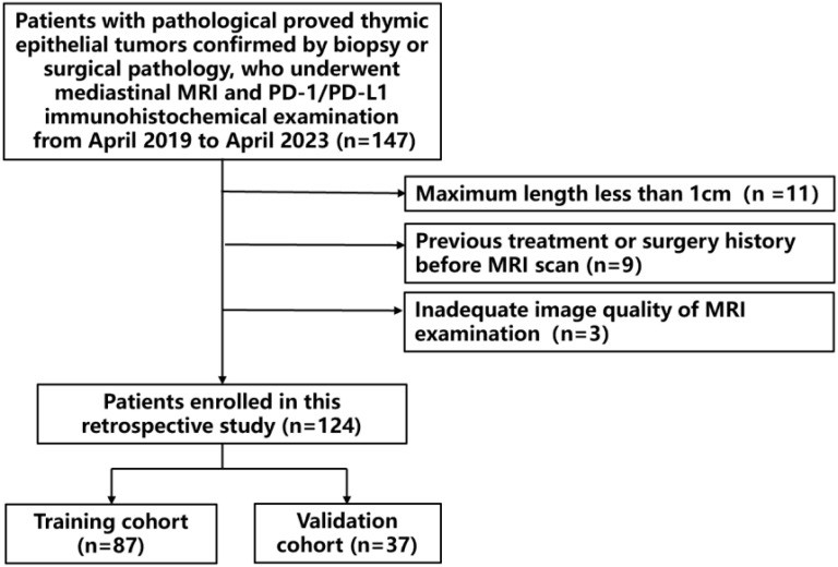 Figure 1
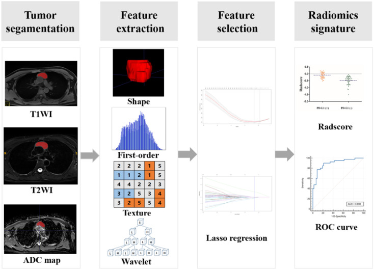 Figure 2
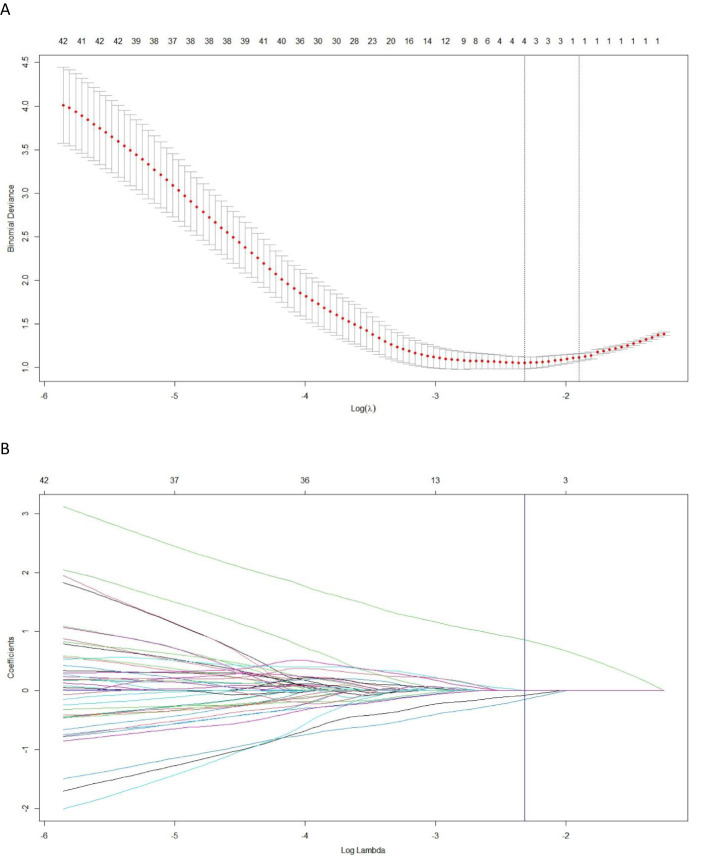 Figure 3
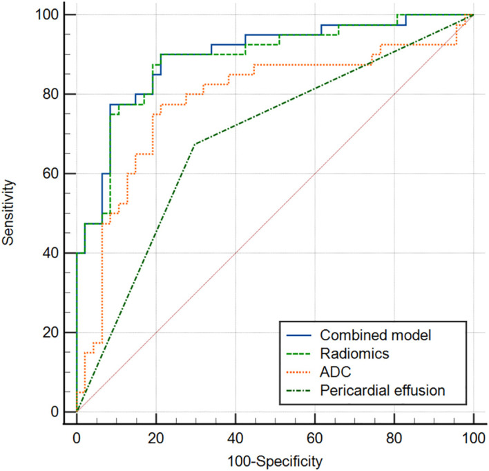 Figure 4
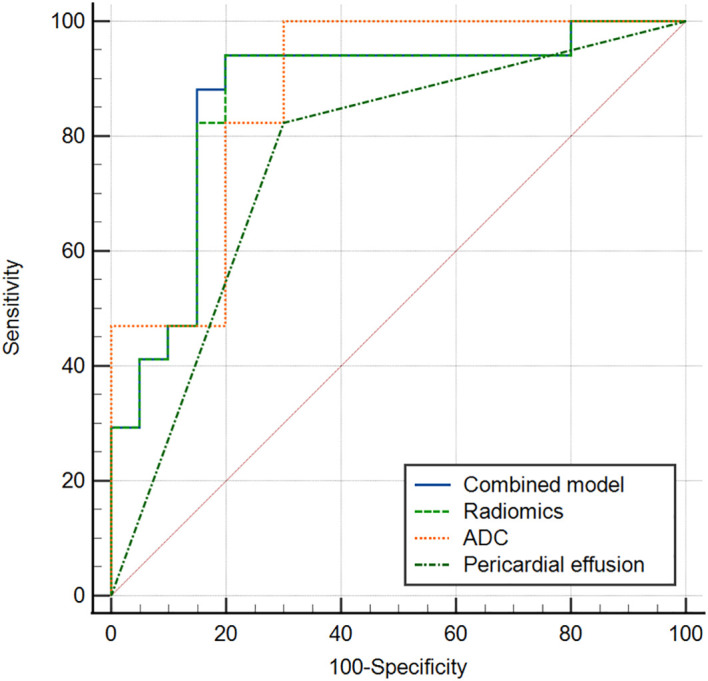 Figure 5
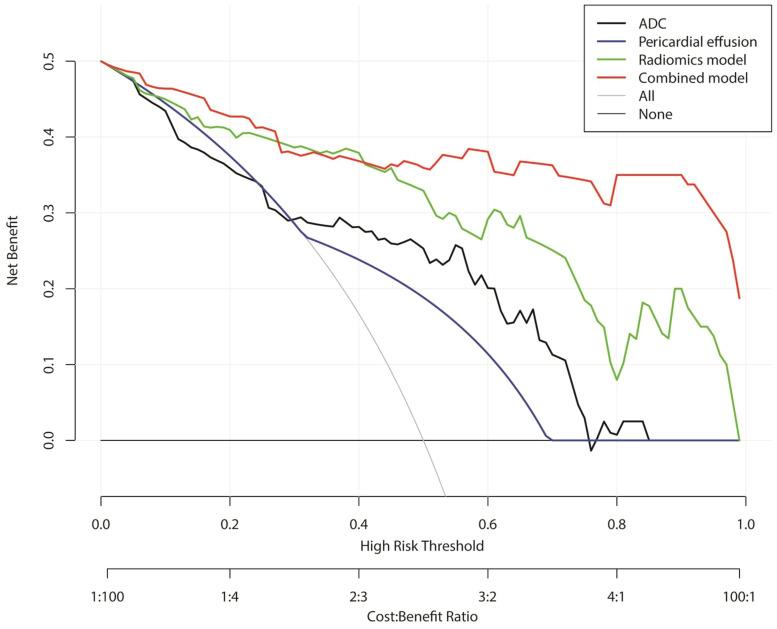 Figure 6
Peer Reviews
No public reviews on file for this paper yet. If you reviewed it on a platform where reviews are public (OpenReview, ICLR, NeurIPS, ICML), you can paste yours below so the community can read it here.
Videos
No videos yet. Explain this paper in a talk, walkthrough, or lecture? Add one.
Taxonomy
TopicsRadiomics and Machine Learning in Medical Imaging · Myasthenia Gravis and Thymoma · Glioma Diagnosis and Treatment