Dimensional changes in buccal cortical bone and lesion volume in teeth with persistent chronic periapical disease subjected to periapical surgery: a cone beam computed tomography study at one year of follow-up
Araceli Boronat-López, Juan Carlos Bernabeu-Mira, Miguel Peñarrocha-Diago, María Peñarrocha-Diago, David Peñarrocha-Oltra

TL;DR
This study used 3D imaging to track changes in jawbone and lesion size after surgery for chronic tooth disease, finding significant reductions in lesion volume over one year.
Contribution
The study provides new insights into healing outcomes after periapical surgery using CBCT, highlighting the role of buccal cortical bone thickness in lesion volume reduction.
Findings
Lesion volume decreased by 91.1% one year after surgery.
Buccal cortical bone thickness predicted smaller volume reduction in lesions.
Anterior teeth showed more significant lesion volume reduction compared to other tooth positions.
Abstract
This study aimed to evaluate changes in buccal cortical bone and lesion volume in teeth with persistent periapical disease one year after periapical surgery using cone-beam computed tomography (CBCT). A prospective study was conducted involving patients with persistent periapical disease undergoing periapical surgery, with one year of follow-up. Data collected included patient age, gender, teeth involved, and the number of roots/lesions. CBCT measurements were taken preoperatively and one year post-surgery, including the distance from the cementoenamel junction to the buccal bone crest (CEJ-BBC), marginal bone loss, buccal cortical height, presence of fenestration, apical depth, cortical bone width at 1, 3, and 5 mm from the buccal bone crest, and lesion volume in mm³. Success was assessed using the “Modified Penn 3D criteria.” The study included 92 patients with 111 roots exhibiting…
Genes, proteins, chemicals, diseases, species, mutations and cell lines named across the full text — each resolved to its canonical identifier and authoritative record.
Click any figure to enlarge with its caption.
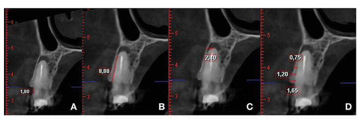 Figure 1
Peer Reviews
No public reviews on file for this paper yet. If you reviewed it on a platform where reviews are public (OpenReview, ICLR, NeurIPS, ICML), you can paste yours below so the community can read it here.
Videos
No videos yet. Explain this paper in a talk, walkthrough, or lecture? Add one.
Taxonomy
TopicsDental Radiography and Imaging · Endodontics and Root Canal Treatments · Sinusitis and nasal conditions