Minimally invasive management of chronic venous insufficiency: A case report on combined radiofrequency ablation and sclerotherapy in an obese patient
M.A. La Marca, S. Bruno, E. Dinoto, R. Federico, F. Pecoraro, D. Mirabella

TL;DR
This case report shows that combining radiofrequency ablation and sclerotherapy is effective for treating chronic venous insufficiency in obese patients.
Contribution
The paper presents a successful case of minimally invasive treatment for CVI in an obese patient with multiple comorbidities.
Findings
Combined radiofrequency ablation and sclerotherapy successfully treated venous insufficiency in an obese patient.
The procedure resulted in no postoperative complications and effective vein obliteration.
The approach is suitable for high-risk patients due to reduced invasiveness and quicker recovery.
Abstract
Chronic venous insufficiency (CVI) affects a significant portion of the population, particularly impacting those with obesity. This condition leads to various symptoms, including leg discomfort and edema, contributing to work absenteeism. Traditional surgical procedures, like saphenous vein stripping and phebectomy, are increasingly supplanted by minimally invasive techniques, such as radiofrequency ablation (RFA) and sclerotherapy, which reduce invasiveness and associated complications, particularly beneficial for high-risk patients, including those with obesity. A 22-year-old male patient with a BMI of 41, suffering from severe varicose veins, hypertension, diabetes, and obstructive sleep apnea. The patient underwent simultaneous RFA and sclerotherapy after imaging confirmed significant venous incompetence but ruled out deep vein thrombosis. The procedure, performed under spinal…
Genes, proteins, chemicals, diseases, species, mutations and cell lines named across the full text — each resolved to its canonical identifier and authoritative record.
Click any figure to enlarge with its caption.
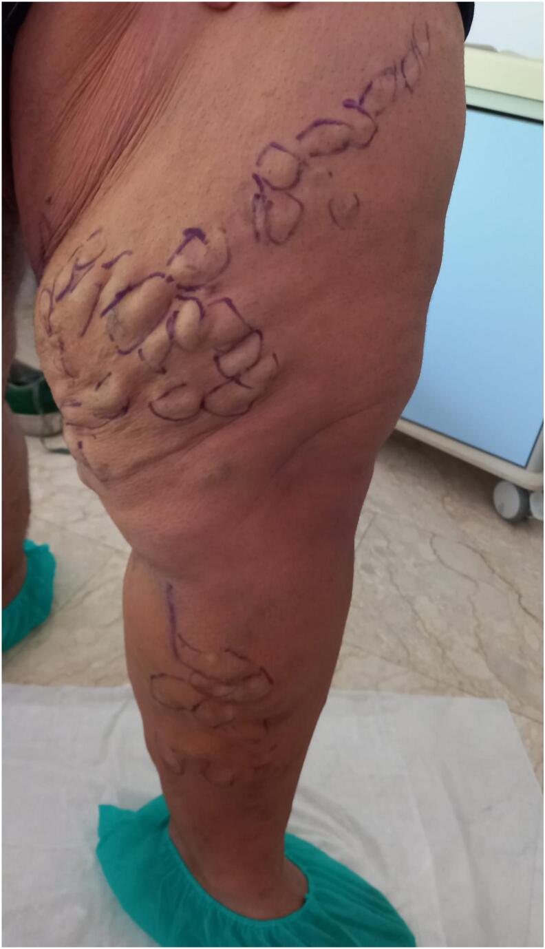 Figure 1
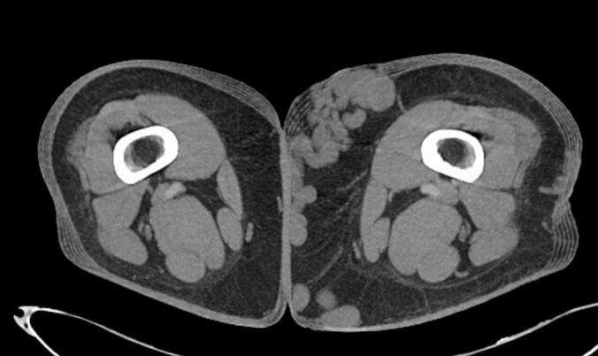 Figure 2
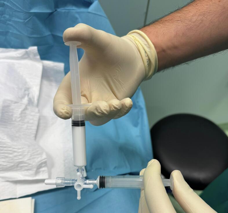 Figure 3
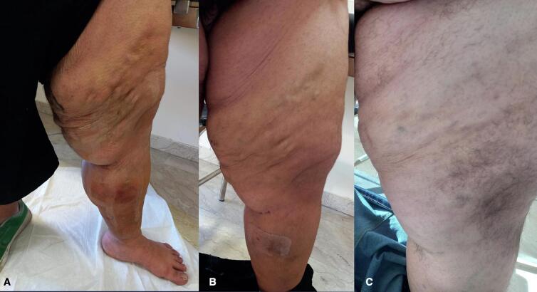 Figure 4
Peer Reviews
No public reviews on file for this paper yet. If you reviewed it on a platform where reviews are public (OpenReview, ICLR, NeurIPS, ICML), you can paste yours below so the community can read it here.
Videos
No videos yet. Explain this paper in a talk, walkthrough, or lecture? Add one.
Taxonomy
TopicsDiagnosis and Treatment of Venous Diseases · Venous Thromboembolism Diagnosis and Management · Dermatologic Treatments and Research
Introduction
1
Chronic venous insufficiency (CVI) affects approximately 10–60 % of population, with an incidence of about 2.6 % in women and 1.9 % in men. It is one of the primary causes of work absenteeism due to symptoms that commonly include leg discomfort, which manifesting as pain, heaviness, itching or burning sensations [1,2]. The main risk factors for CVI include age, gender, family history, and prolonged periods of sedentary or standing posture [3]. The issue of venous insufficiency is particularly significant in obese patients, who face unique challenges in both a surgical interventions and the management of pharmacological and elastic compression therapies [4]. According to the Clinical, Etiological, Anatomical, and Physiopathological (CEAP) classification, venous insufficiency can have primary, secondary, or congenital etiology (Table 1). The most common primary etiology is the loss of elasticity in the venous wall, which leads to the incompetence of unidirectional valves and subsequently causes venous hypertension. It is essential to consider post-thrombotic syndromes and congenital alterations, as superficial venous insufficiency may be an epiphenomenon that conceals more serious underlying conditions that should be addressed in therapy planning [5,6]. CVI is characterized by various stages, ranging from asymptomatic conditions, primarily aesthetic in nature, to severe, debilitating states with ulcerations that can affect large areas of skin tissue. [7] Traditional procedures such as saphenous vein stripping and phlebectomy are increasingly being supplanted by minimally invasive techniques, including Endovenous Laser Ablation (EVLA) and Radiofrequency Ablation (RFA), often in conjunction with sclerotherapy. This shift aims to minimize the invasiveness and complications associated with open surgery. Furthermore, these techniques can often be performed in a single session, making them suitable for patients who may not be candidates for conventional open surgery, such as those with obesity. In this report, we present a case of simultaneous treatment utilizing RFA and sclerotherapy in a patient with severe varicose veins and high-grade obesity (Body Mass Index >35). This work has been written in accordance with the SCARE criteria [8,9].Table 1CEAP classification.Table 1. ClinicalC ClassDescriptionC0No visible or palpable signs of venous diseaseC1Teleangectasieas or reticular veinsC2Varicose VeinsC2rRecurrent Varicose VeinsC3EdemaC4Changes in Skin and subcutaneous Tissue Secondary to Chronic Vein DiseaseC4aPigmentation or EczemaC4bLpodermatosclerosis or Atrophie BlancheC4cCorona PhebectaticaC5Healed Venous UlcerC6Active Venous UlcerC6rRecurrent Active Venous Ulver EtiologicalE ClassDescriptionEpPrimaryEsSecondaryEsiSecondary - IntravenousEseSecondary - ExtravenousEcCongenitalEnNo Cause Identified AnatomicalA ClassDescriptionAsSuperficialDescriptionOldNewTeleangectasiea1TelReticular Veins1RetGreat Saphenous Vein Above Knee2GSVaGreat Saphenous Vein Belove Knee3GSVbSmall Saphenous Vein4SSVAnterior Accessory Saphenous Vein4AASVNonsaphenous Vein5NSVAdDeepInferior Vena Cava6IVCCommon Iliac Vein7CIVInternal ciac Vein8IIVExternal Iliac Vein9EIVPelvic Veins10PELVCommon Femoral Vein11CFVDeep Femoral Vein12DFVFemoral Vein13FVPopliteal Vein14POPVCrural (Tibial) Vein15TIBVMuscular Vein16MUSVGastrocnemius Vein16GAVSoleal Vein16SOVPerforatorThigh Perforator Vein17TPVCalf Perforator Vein18CPVAnNo Venous Anatomic Location Identified PathophysiologicalP ClassDescriptionPrRefluxPoObstructionPr,oReflux and ObstructionPnNo pathophysiology Identified
Case report
2
A 22-year-old male patient with obesity, characterized by a Body Mass Index (BMI) of 41, presents with a variety of comorbid conditions, including hypertension, diabetes, obstructive sleep apnoea syndrome (OSAS), and anxiety-depressive disorder. He reports pain and a sensation of heaviness in his left lower limb, accompanied by significant edema (CEAP C4a) (Fig. 1). The patient denies any history of prior surgeries. Due to the patient's severe obesity, the results of the ultrasonography were inconclusive regarding the assessment of the deep venous system; however, they did indicate significant valvular incompetence at the saphenofemoral junction, extending to the distal third of the thigh along the great saphenous vein (GSV). To exclude the possibility of deep vein thrombosis (DVT) or other abnormalities that might contribute to venous hypertension, a computed tomography angiography (CTA) of the abdomen and lower limbs was performed. This imaging ruled out deep veins involvement and confirmed the presence of multiple venous ectasias affecting the superficial venous system (Fig. 2). The ectasias were most prominent in the soft tissues beginning at the anterolateral root of the left thigh and extending along the medial and posterior aspects of the middle third of the thigh, as well as the anteromedial side of the middle third of the leg. The extensive network of venous branches posed significant challenges for the treatment of varicose veins; any phlebectomies would require multiple incisions, therapy increasing the risk of infection. Given the diameters of the saphenous vein (maximum diameter of 8.9 mm in the distal third of the thigh and 15.7 mm at the saphenofemoral junction) and its relatively straight course, the patient was identified as a suitable candidate for saphenectomy, combined with RFA and sclerotherapy of the collateral veins. Taking into account the patient's size, limited cooperation due to anxiety-depressive syndrome, and the OSAS which could heighten the risk associated with increased intraoperative sedation, it was determined that performing the procedure under spinal anesthesia would be preferable. A 7 French introducer was inserted at the proximal third of the left leg. A radiofrequency device (vienCLEAR - RF Medical, Seoul, Korea) was introduced and positioned 2.5 cm from the saphenofemoral junction. To facilitate the adherence of the probe to the walls of the GSV, a tumescent solution totaling 250 ml was infused along the entire length of the vein. After ultrasound-guided confirmation of the correct placement of the guide tip, the GSV was ablated. The catheter was then connected to a generator that delivers radiofrequency energy upon activation. Each 7-cm segment of the vein was heated for 20 s before the energy supply automatically shut off. The catheter was then manually advanced to the next marker, and the heating process was repeated for subsequent segments. The proximal 7-cm segment of the GSV underwent two 20-s energy cycles, while the other segments were treated with either one or two cycles. For each 20-s cycle, the temperature required to reach 120 °C within 5 s of activation. If this temperature was not achieved within that timeframe, the segment underwent an additional 20-s treatment cycle. Post-ablation ultrasound check confirmed the complete obliteration of the GSV while ensuring the patency of some collateral veins responsible for extra-saphenous varicosities. The introducer was subsequently removed. To prevent any potential early recanalizations of the GSV due to the permeability of the saphenous collaterals, treatment continued with the Ultrasound-guided injection of 3 ml of 1 % Lauromacrogol foam into the venous collaterals of the proximal third of the leg (1 ml) and the middle third of the thigh (2 ml), followed by a dressing and elastic compression bandage (Fig. 3). The postoperative course was smooth, with the patient reporting no significant pain or swelling and 1 g paracetamol tablets were administered as needed. The patient was discharged home the following day. Follow-up appointments were scheduled at 15 days, 1 month and 6 months post-procedure, and ultrasound assessments indicated no complications or recurrences, allowing the patient to return to work without symptoms (Fig. 4).Fig. 1. Varicose veins in obesity patient before procedure.Fig. 1. Fig. 2CT scan with widespread and significant varicose veins.Fig. 2. Fig. 3Preparation of foam 1 % Lauromacrogol.Fig. 3. Fig. 4Outcome at 15 days (A), one month (B), 6 months (C).Fig. 4
Discussion
3
The prevalence of lower limb venous diseases and obesity is increasing. Numerous studies have shown that being overweight is a contributing factor to CVI, which can lead to the early onset of ulcerations. [[10], [11], [12]]. Surgical interventions on the superficial venous system typically carry a low operative risk; however, a patient's comorbidities can elevate the risks associated with these procedures. High ligation and stripping of GSV are usually conducted in an operating room under spinal or general anesthesia. Whenever possible, major forms of anesthesia should be avoided to reduce complications related to anesthetics, such as allergic reactions, damage to teeth during intubation, and post-operative nausea and vomiting [13]. Currently, treatment options for CVI include endovascular techniques such as RFA and EVLA, sclerotherapy and surgical therapies. Studies have demonstrated the effectiveness and safety of endovascular procedure compared with traditional open treatment [14]. The Society for Vascular Surgery and the American Venous Forum recommend endovascular techniques as the primary choice for treating superficial venous disease due to their reduced pain, lower morbidity, shorter hospital stays and quicker return to work [15]. The literature indicates that obesity is a significant risk factor for complications associated with any type of surgical intervention due to increased risks of cardiovascular, respiratory, and site infections [16,17]. Obese individuals are 47 % more likely to experience CVI compared to those with a normal body mass index. Previous research has indicated that changes in venous biomechanics, resulting from venous return obstruction linked to increased abdominal adipose tissue, contribute to a higher risk of cardiovascular disease in obese patients. Additionally, individuals with obesity often struggle with adhering to compression therapy due to difficulties in donning or wearing compression stockings, which may be influenced their body habitus. This can lead to poor compliance with compression therapy and diminish the clinical response following venous treatments. Moreover, it has been shown that BMI adversely affects the CEAP clinical classification, pain levels, and quality of life, independently of venous reflux. BMI has also been identified as a significant factor associated with less improvement in the revised Venous Clinical Severity Score after venous treatment [18]. Rodriguez-Avecedo et al. identified a BMI >30 as a potential criterion for distinguishing between patients at increased risk of recanalization and those who are not [19]. Other causes of early recanalization, as reported by Bissaco et al., may be attributed to specific sources of reflux, such as anterior or posterior accessory saphenous veins and perforating veins, irrespective of the primary ablation treatment [20]. Consequently, opting for a less invasive technique seems a logical choice, especially in overcoming the potential challenges posed by open techniques. The combination of multiple minimally invasive techniques may enhance the success rate and sustainability of treatment outcomes [21]. Ultrasound-guided foam sclerotherapy, combined with RFA closure, represents a novel approach to treating CVI [22]. This technique generates thermal energy via a radiofrequency catheter, targeting the intima and collagen fibers of affected venous vessels. The resulting thermal coagulation effect damages endothelial cells, leading to degeneration, thickening, organization, contraction, and shedding [23]. This process helps narrow the venous lumen and thicken the venous wall, ultimately closing varicose veins through the formation of fibrotic cords and achieving the desired therapeutic outcome [24]. The ultrasound-guided sclerotherapy, performed during the thermal ablation procedure, effectively eliminates the presence of feeding collateral veins that could supply the saphenous vein, one of the risk factors for recanalization. This minimally invasive procedure does not require stripping to eliminate the diseased venous vessels; instead, it occludes venous blood flow under local anesthesia to produce the therapeutic effect. This approach alleviates pain and minimizes associated complications, making it more acceptable to patients [25]. Numerous positive experiences have emerged from the combination of these two techniques. Zhao and Hongtao, in separate studies, demonstrated that the quality of life for patients treated with a combination technique was significantly higher than that of the control group six months after the operation (P < .05). This suggests that ultrasound-guided foam sclerotherapy combined with intracavity radiofrequency closure can effectively enhance the quality of life for patients with CVI by reducing the procedure length, postoperative bed rest time, hospital stays, and intraoperative blood loss when compared to traditional open treatment methods [21,26]. Memon et al. treated 102 cases with radiofrequency, of which 79 % underwent concurrent sclerotherapy. During the follow-up, 99 % of the treated patients experienced a permanent vessel occlusion [27]. Carrol et al. highlight in a literature review that out of 1453 unique citations, including 34 randomized clinical trials (54 articles), minimally invasive techniques yielded clinical results comparable to traditional surgery. Recurrence rates were slightly lower for EVLA, RFA, and Foam Sclerotherapy, especially during longer follow-up periods. Short-term pain was lower for Foam Sclerotherapy and RFA but higher for EVLA. All evaluated techniques reported better quality of life scores compared to stripping. Therefore, the differences between treatments were found to be negligible in terms of clinical outcomes, suggesting that the most cost -effective treatment is the one with the lowest cost. The total costs of minimally invasive treatments were lower and marginally more effective compared to stripping [28]. Among the limited studies focused on obese patients, Zottola et al. reported that those in higher obesity classes experience greater reductions in perceived symptoms during early follow-up after minimally invasive interventions. While obesity has been associated with increased severity of venous disease symptoms, obese patients can experience significant short-term relief following treatment, often achieving greater symptom improvement compared to non-obese patients, particularly when treated for milder disease presentations [12]. The methodological limitations are evident in the contraindications associated with these procedures. For instance, excessive tortuosity of the GSV can complicate RFA, while allergies to sclerosing agent can hinder sclerotherapy. Unfortunately, many pivotal studies have specifically excluded obese individuals [29]. Nevertheless, the substantial presence of obese patients suffering from venous insufficiency highlights the need for effective and potentially repeatable treatments. In our experience, the application of a combined technique in high-risk patient for complications and recurrence has allowed us to achieve the desired outcomes while minimizing risks.
Conclusions
4
Obesity is identified as a considerable risk factor for complications related to surgical interventions for CVI. The combination of RFA and simultaneous sclerotherapy is a technique that has shown a high rate of technical success and an improvement in postoperative quality of life. These findings emphasize the importance of customizing treatment strategies to align with individual patient profiles, especially for those who are obese or have other comorbidities. This customization is essential to ensure effective management of CVI while minimizing the risk of complications. Further research is needed to explore the long-term outcomes of combined minimally invasive techniques for CVI in obese patients, as existing literature frequently overlooks this demographic. The demand for accessible and effective treatment options for this population remains a critical issue in managing CVI.
Credit authorship contribution statement
Conceptualization, MA.L., E.D. and D.M.; methodology, E.D., R.F.; software, M.A.L; validation, D.M., E.D. and F.P.; formal analysis, S.B. and E.D.; investigation, M.A.L.; resources, M.A.L., R.F.; data curation, E.D., D.M.; writing—original draft preparation, M.A.L.; writing—review and editing, E.D., D.M.; visualization, D.M., F.P.; supervision, F.P.; project administration, F.P. All authors have read and agreed to the published version of the manuscript.
Informed consent statement
Written informed consent was obtained from the patient for publication of this case report and accompanying images. A copy of the written consent is available for review by the Editor-in-Chief of this journal on request.
Ethical approval
All procedures followed were in accordance with the ethical standards of the Institutional Committee on Human Experimentation and with the Helsinki Declaration. According to the internal review board, the retrospective and anonymized nature of the study did not require medical ethical committee approval. This article does not require ethics committee approval due to the retrospective nature of the paper and the type of technique reported.
Institutional review board statement
All procedures followed were in accordance with the ethical standards of the Institutional Committee on Human Experimentation and with the Helsinki Declaration. According to the internal review board, the retrospective and anonymized nature of the study did not require medical ethical committee approval.
Guarantor
ETTORE DINOTO.
Funding
This research received no external funding.
Declaration of competing interest
The authors declare no conflict of interest.
The reference list from the paper itself. Each links out to its DOI / PubMed record.
- 1Qari T.A.Almatrafi K.N.Khateb F.R.Al-Kaabi B.Al-Harbi A.Alabdali S.Al-Nemari R.Bannani S.A.Prevalence of varicose veins among surgeons: a cross-sectional study Cureus 1682024 Aug 24e 6768710.7759/cureus.67687 PMID: 39314592; PMCID: PMC 11419730 PMC 1141973039314592 · doi ↗ · pubmed ↗
- 2Al Shammeri O.Al Hamdan N.Al-Hothaly B.Midhet F.Hussain M.Al-Mohaimeed A.Chronic venous insufficiency: prevalence and effect of compression stockings Int. J. Health Sci. (Qassim)832014 Jul 23123610.12816/0023975 PMID: 25505858; PMCID: PMC 425735825505858 PMC 4257358 · doi ↗ · pubmed ↗
- 3Ortega M.A.Fraile-Martínez O.García-Montero C.Álvarez-Mon M.A.Chaowen C.Ruiz-Grande F.Pekarek L.Monserrat J.Asúnsolo A.García-Honduvilla N.Álvarez-Mon M.Bujan J.Understanding chronic venous disease: a critical overview of its pathophysiology and medical management J. Clin. Med.10152021 Jul 22323910.3390/jcm 10153239 PMID: 34362022; PMCID: PMC 834867334362022 PMC 8348673 · doi ↗ · pubmed ↗
- 4Popplewell M.A.Mahesh S.Nandra S.Juszczak M.Ashby H.Wall M.L.The obese population’s views on the symptoms and risks of chronic venous insufficiency - 2 (OBVIOUS-2) cross-sectional survey Phlebology 2024 Sep 1710.1177/026835552412841792683555241284179. (Epub ahead of print. PMID: 39287433)PMC 1195136139287433 · doi ↗ · pubmed ↗
- 5Raetz J.Wilson M.Collins K.Varicose veins: diagnosis and treatment Am. Fam. Physician 99112019 Jun 1682688(PMID: 31150188)31150188 · pubmed ↗
- 6Raffetto J.D.Khalil R.A.Mechanisms of Lower Extremity Vein Dysfunction in Chronic Venous Disease and Implications in Management of Varicose Veins. Vessel Plus 520213610.20517/2574-1209.2021.16Epub 2021 May 29. PMID: 34250453; PMCID: PMC 8270011 PMC 827001134250453 · doi ↗ · pubmed ↗
- 7Atay M.Altun S.Outcomes of radiofrequency ablation therapy of great saphenous veins insufficiency J. Coll. Physicians Surg. Pak.3282022 Aug 1009101310.29271/jcpsp.2022.08.1009 PMID: 3593212435932124 · doi ↗ · pubmed ↗
- 8Agha R.A.Borrelli M.R.Farwana R.Koshy K.Fowler A.J.Orgill D.P.PROCESS Group. The PROCESS 2018 statement: Updating Consensus Preferred Reporting Of Cas E Series in Surgery (PROCESS) guidelines Int. J. Surg.602018 Dec 27928210.1016/j.ijsu.2018.10.031Epub 2018 Oct 22. PMID: 3035978130359781 · doi ↗ · pubmed ↗