“Neuropathological function estimations”: a user-friendly module for analyzing neural activity in neurological disorders
Alessia M Panait, Alex Kim, Rafael Rodriguez-Rojas, Lazaro M Sanchez-Rodriguez, Yasser Iturria-Medina

TL;DR
A new software tool helps researchers study brain activity changes in neurological disorders without needing programming skills.
Contribution
The tool introduces a user-friendly platform for generating personalized brain models and analyzing neuropathological influences.
Findings
The software was validated using Alzheimer’s disease data to study neuronal excitability.
It provides insights into disease mechanisms and supports personalized therapeutic research.
Abstract
This work introduces the Neuropathological Function Estimations software, designed to facilitate the study of neuronal activity alterations in neurological disorders without requiring programming expertise. With its user-friendly interface, researchers can input various data types to generate subject-specific functional brain models and decode neuropathological influences. The software’s capabilities are validated through its application to Alzheimer’s disease, providing insights into neuronal excitability and disease mechanisms. This tool has the potential to enhance our understanding of the biological basis of in vivo neural activity and contribute to the development of personalized therapeutic interventions. The latest version of the software and support are freely available for noncommercial users through the Neuroinformatics for Personalized Medicine Lab (NeuroPM Lab) website at…
Genes, proteins, chemicals, diseases, species, mutations and cell lines named across the full text — each resolved to its canonical identifier and authoritative record.
Click any figure to enlarge with its caption.
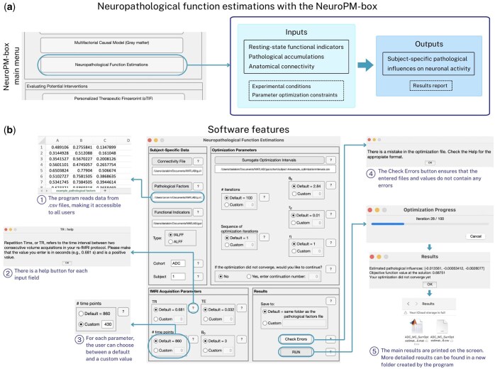 Figure 1
- —Natural Sciences and Engineering Research Council of Canada10.13039/501100000038
- —Canadian Institutes of Health Research10.13039/501100000024
Peer Reviews
No public reviews on file for this paper yet. If you reviewed it on a platform where reviews are public (OpenReview, ICLR, NeurIPS, ICML), you can paste yours below so the community can read it here.
Videos
No videos yet. Explain this paper in a talk, walkthrough, or lecture? Add one.
Taxonomy
TopicsFunctional Brain Connectivity Studies
1 Introduction
Identifying the biological basis of neural activity abnormalities is instrumental for understanding brain disease (Sanchez-Rodriguez et al. 2024a,b). Since direct in vivo measurements of neuronal dysfunction often require invasive techniques, computational models have been developed as an alternative for studying neuronal activity in humans (Sanchez-Rodriguez et al. 2024a). Utilizing these models requires advanced programming and biophysical modeling expertise, however. Furthermore, while tools like The Virtual Brain (Schirner et al. 2022) serve as references for full-brain simulations, they currently lack built-in capabilities that can be widely applied to estimate biophysical alterations due to disease. To further democratize the characterization of causal pathological effects on neuronal activity in neurological disorders, we implemented a new module into the NeuroPM-box (Iturria-Medina et al. 2021). This user-friendly toolbox integrates diverse data to create realistic brain models and identify optimal personalized treatments.
2 Neuronal activity changes: core mechanisms in neurological disorders
Neural networks rely on balanced activity between excitatory and inhibitory neurons, which is essential for normal brain functioning. Disruptions in neuronal activity have been observed in many neurological disorders and are increasingly regarded as key mechanistic events (Ghatak et al. 2021). For instance, epileptogenesis often manifests as neuronal hyperexcitability and hypersynchrony (Chen et al. 2017). In Parkinson’s disease, dopamine depletion alters activity in the basal ganglia-thalamocortical network, which is responsible for the observed cardinal motor features (Foffani et al. 2019). In Alzheimer’s disease (AD), amyloid-beta (Aβ) plaques and tau tangles physically impair neurons, and their complex interactions seem to increase neuronal excitability, correlating with cognitive performance and several disease biomarkers (Sanchez-Rodriguez et al. 2024a).
We previously developed a biophysical framework to characterize neuronal activity alterations in AD (Sanchez-Rodriguez et al. 2024a). Aβ and tau accumulations in a brain region, observed in the subject’s PET scans, were assumed to linearly influence a neuronal excitability parameter, spreading across brain regions through connections identified via diffusion MRI. The model then simulated pathophysiological brain activity patterns and converted them into resting-state fMRI signals. Finally, the effects of Aβ and tau were quantified by fitting the generated signals to the subject’s real fMRI signals in terms of their fractional amplitude of low-frequency fluctuations (fALFF) (Jia et al. 2019), a reliable marker for neuronal activity (Supplementary Fig. S1). In this work, we extend the code applied to AD, rendering it more accessible, user-friendly, and easily adaptable for studying other conditions.
3 Software description
The software was designed to accelerate the investigation of neuronal activity alterations in neurological disorders. This graphical user interface, named “Neuropathological Function Estimations,” was developed in MATLAB R2024a (The MathWorks Inc., Natick, MA, USA). It is available as a standalone application for the major operating systems: Windows, macOS, and Linux, and requires the installation of MATLAB Runtime to function. The expected processing time varies depending on the operating system and device model, and on average it takes about an hour to complete 100 iterations of the optimization algorithm (see Supplementary Text S2: Instructions for installation and use). Researchers can upload their data in .csv format, which is widely accessible. The software requires four main inputs for each subject: (i) brain-wide distributions of the pathological factors included in the model, (ii) non-invasive in vivo indicators of functional alterations (typically obtained through resting-state fMRI), (iii) anatomical connectivity values for the regions defined in the user’s brain parcellation, and (iv) experimental conditions and parameter optimization constraints (see Fig. 1a and the exemplary simulated data provided with the software). Using this information, a whole-brain neuropathological influence model is generated within the code, and the most likely neuropathological influence parameters are identified through a surrogate optimization algorithm (https://www.mathworks.com/help/gads/surrogateopt.html).
Functionalities of the Neuropathological Function Estimations module. (a) The software processes regional inputs including resting-state functional indicators and pathological accumulations, inter-regional anatomical connectivity, and experimental conditions/parameter optimization constraints. Consequently, the software identifies subject-specific pathological influences on neuronal activity and generates a comprehensive results report. (b) The tool is designed to be highly user-friendly. The main features that enhance the user experience have been highlighted in the figure. Default parameters and options are based on previous publications related to Alzheimer’s disease (Sanchez-Rodriguez et al. 2024a).
The Neuropathological Function Estimations software features extensive personalization options and a clear layout, as shown in Fig. 1b. Each button group offers a default option with suggested standard values from the literature, as well as a custom option that allows users to specify their own parameters. Help texts are conveniently provided next to each parameter and button. Additionally, a complete step-by-step guide for utilizing the software, detailed installation instructions and mathematical formulations are included in the Supplementary Material. The software consists of several key sections. In the Subject-Specific Data section, users can input files containing biological measurements for each participant, select the type of functional indicators used, and tag their cohort and individual participants. Users can specify the parameters of their imaging protocol in the fMRI Acquisition Parameters section. The program will then generate resting-state fMRI signals according to these provided values. The Optimization Parameters section offers options regarding the optimization methods used to calculate results. For instance, users can choose the number of iterations for the run and decide whether to continue from a previous calculation that did not converge. Finally, the Results section includes options for selecting the file save location, as well as a “Check Errors” button to verify that the files and values entered do not contain any errors. Pressing the “RUN” button initiates the calculations. Once all calculations are complete, .mat and .csv files containing the outputs are generated and also appear in a pop-up window on the screen. The software has been tested on benchmark data, showing excellent alignment (average R^2^ = 0.97) between ground-truth and reconstructed neuronal activity parameter distributions (see Supplementary Fig. S2).
One significant limitation is that the current implementation is computationally intensive due to the simulations of the subject’s resting-state fMRI signals. However, acceleration can be achieved by reducing the number of time points used to calculate the functional indicators, without sacrificing biological reliability (Küblböck et al. 2014). Moreover, our model is based on the assumption that neuronal excitability can be reliably estimated by a simplified model of connected excitatory and inhibitory populations, which may pose a challenge in capturing the complexity of the living brain. Additionally, to ensure that the dynamical systems produce oscillatory behavior, it is recommended that users begin by applying narrow optimization intervals—see the discussion in (Sanchez-Rodriguez et al. 2024a) and the references therein for more information on the biophysical parameters of the model. The obtained values and comparisons among the subject-specific estimated pathophysiological influences help identify the molecular mechanisms underlying neuronal dysfunction and inform potential therapeutic interventions, among other possibilities (Sanchez-Rodriguez et al. 2024a,b).
4 Conclusion
The software reconstructs key biological quantities of interest and enhances our understanding of network reorganization patterns in the brain and their relationship with disease symptoms and prognosis. In the case of AD, we have validated the effectiveness of our computational approach in disease hypothesis testing (Sanchez-Rodriguez et al. 2024a,b), expanding the capabilities of the existing ecosystem of large-scale brain network simulations and parameter identification platforms. For example, the latest version of The Virtual Brain simulator (Schirner et al. 2022) enables Bayesian parameter optimization in epilepsy patient models. Our tool paves the way for new applications in other diseases that also cause neuronal activity alterations and complements the robust suite of methods for multifactorial disease analysis already available in the NeuroPM-box (Iturria-Medina et al. 2021). For instance, in Parkinson’s disease, dopamine transporter imaging techniques such as DaT 123I–FP-CIT scans and alpha-synuclein-specific PET (once available) can provide insights into the disease’s pathology, while fALFF and ALFF may serve as non-invasive, in vivo indicators of functional alterations (Lin et al. 2022, Khan et al. 2023, Sanchez-Rodriguez et al. 2024a). Future releases will offer greater flexibility in selecting affected parameters and neuronal activity markers, including additional resting-state fMRI or E/MEG features, as well as the ability to define different pathological influence equations. We also plan to add new features to handle increasing data volumes (e.g. finer brain parcellations requiring more physical memory to run the dynamical equations). An upcoming article describes an application that ensures scalability by using machine learning algorithms to predict functional indicators for any parameter combination from just a few grid simulations, reducing the need for ad-hoc runs. These ongoing efforts to refine the computational pipeline aim to improve both the efficiency and precision of the modeling approach. Understanding the mechanisms behind disease manifestations is crucial for developing personalized, effective therapeutic interventions.
Supplementary Material
vbaf083_Supplementary_Data
The reference list from the paper itself. Each links out to its DOI / PubMed record.
- 1Chen Z , An Y, Zhao B et al The value of resting-state functional magnetic resonance imaging for detecting epileptogenic zones in patients with focal epilepsy. P Lo S One 2017;12:e 0172094.28199371 10.1371/journal.pone.0172094 PMC 5310782 · doi ↗ · pubmed ↗
- 2Foffani G , Trigo-Damas I, Pineda-Pardo JA et al Focused ultrasound in Parkinson’s disease: a twofold path toward disease modification. Mov Disord 2019;34:1262–73.31412430 10.1002/mds.27805 · doi ↗ · pubmed ↗
- 3Ghatak S , Talantova M, Mc Kercher SR et al Novel therapeutic approach for excitatory/inhibitory imbalance in neurodevelopmental and neurodegenerative diseases. Annu Rev Pharmacol Toxicol 2021;61:701–21.32997602 10.1146/annurev-pharmtox-032320-015420 PMC 13138087 · doi ↗ · pubmed ↗
- 4Iturria-Medina Y , Carbonell F, Assadi A et al Integrating molecular, histopathological, neuroimaging and clinical neuroscience data with Neuro PM-box. Commun Biol 2021;4:614.34021244 10.1038/s 42003-021-02133-x PMC 8140107 · doi ↗ · pubmed ↗
- 5Jia X-Z , Wang J, Sun H-Y et al RES Tplus: an improved toolkit for resting-state functional magnetic resonance imaging data processing. Sci Bull (Beijing) 2019;64:953–4.36659803 10.1016/j.scib.2019.05.008 · doi ↗ · pubmed ↗
- 6Khan AF , Adewale Q, Lin S-J et al Patient-specific models link neurotransmitter receptor mechanisms with motor and visuospatial axes of Parkinson’s disease. Nat Commun 2023;14:6009.37752107 10.1038/s 41467-023-41677-w PMC 10522603 · doi ↗ · pubmed ↗
- 7Küblböck M , Woletz M, Höflich A et al Stability of low-frequency fluctuation amplitudes in prolonged resting-state f MRI. Neuroimage 2014;103:249–57.25251869 10.1016/j.neuroimage.2014.09.038 · doi ↗ · pubmed ↗
- 8Lin S-J , Rodriguez-Rojas R, Baumeister TR et al Neuroimaging signatures predicting motor improvement to focused ultrasound subthalamotomy in Parkinson’s disease. NPJ Parkinsons Dis 2022;8:70.35665753 10.1038/s 41531-022-00332-9PMC 9166695 · doi ↗ · pubmed ↗