Dynamic and Static Functional Gradient in Temporal Lobe Epilepsy With Hippocampal Sclerosis Versus Healthy Controls
Kangrun Wang, JiaYao Li, Fangfang Xie, Chaorong Liu, Langzi Tan, Jialinzi He, Xianghe Liu, Ge Wang, Min Zhang, Haiyun Tang, Danlei Wei, Jingwan Feng, Sha Huang, Jinxin Peng, Zhuanyi Yang, Xiaoyan Long, Bo Xiao, Juan Li, Lili Long

TL;DR
This study compares brain connectivity patterns in people with temporal lobe epilepsy and healthy individuals, finding disrupted brain gradients linked to cognitive decline.
Contribution
The paper introduces a novel analysis of dynamic and static brain gradients in TLE patients with hippocampal sclerosis.
Findings
TLE patients show lower dynamic recruitment of brain gradients compared to healthy controls.
Atypical activation-gradient correlations in TLE are linked to cognitive impairment.
Principal gradient reconfiguration is not driving activation reorganization in TLE.
Abstract
The gradient captures the continuous transitions in connectivity, representing an intrinsic hierarchical architecture of the brain. Previous works hinted at the dynamics of the gradient but did not verify them. Cognitive impairment is a common comorbidity of temporal lobe epilepsy (TLE). Gradient techniques provide a framework that could promote the understanding of the neural correlations of cognitive decline. Thirty patients with TLE and hippocampal sclerosis and 29 matched healthy controls (HC) were investigated with verbal fluency task‐based functional MRI and gradient techniques. The correlation between task‐based activation/deactivation and healthy gradients, task‐based gradients, and dynamic features calculated with sliding window approaches was compared between HC and TLE. The allegiance in the real data of HC and TLE was more widespread compared to static null models. TLE has…
Genes, proteins, chemicals, diseases, species, mutations and cell lines named across the full text — each resolved to its canonical identifier and authoritative record.
Click any figure to enlarge with its caption.
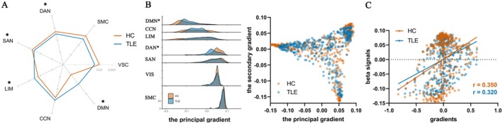 Figure 1
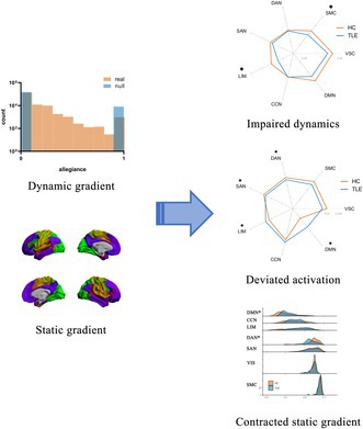 Figure 2
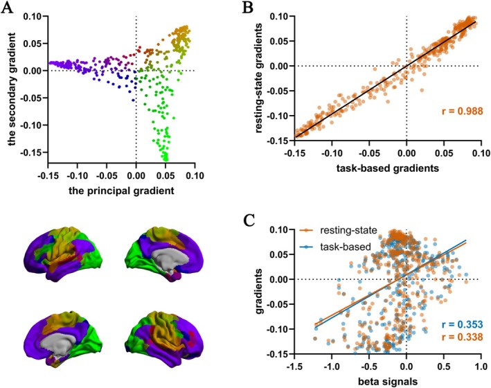 Figure 3
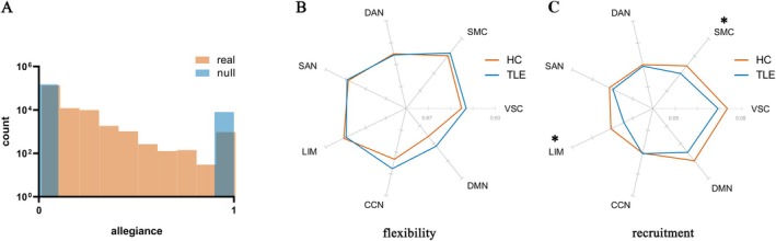 Figure 4
Peer Reviews
No public reviews on file for this paper yet. If you reviewed it on a platform where reviews are public (OpenReview, ICLR, NeurIPS, ICML), you can paste yours below so the community can read it here.
Videos
No videos yet. Explain this paper in a talk, walkthrough, or lecture? Add one.
Taxonomy
TopicsFunctional Brain Connectivity Studies · Advanced MRI Techniques and Applications · Advanced Neuroimaging Techniques and Applications