Micro-CT and histological assessment of renal arterial embolization with Glubran®2 cyanoacrylate: a medium-term follow-up study in a rabbit model
Romaric Loffroy, Kévin Guillen, Olivier Chevallier, Mohamed Fouad, Emilie Couloumy, Anne Dencausse, Philippe Robert, Sarah Catoen, Anne-Virginie Salsac, Serge Ludwig Aho-Glele, Pierre-Olivier Comby

TL;DR
This study evaluates the effectiveness of Glubran®2 glue for kidney artery embolization in rabbits, finding that it maintains occlusion despite partial glue resorption.
Contribution
The study introduces micro-CT as a valuable tool for assessing embolic cast changes and confirms Glubran®2's occlusion durability in a rabbit model.
Findings
Glubran®2 glue maintained arterial occlusion despite partial cast resorption and fragmentation.
Compensatory neovascularization was observed, with no significant inflammation differences between glue concentrations.
Micro-CT scans effectively tracked glue cast changes over time.
Abstract
Cyanoacrylate glues are widely used in interventional radiology as effective embolic agents due to their rapid polymerization and ability to achieve vessel occlusion. Nonetheless, concern remains regarding cast stability and potential recanalization over time. This study used multiple modalities to evaluate the medium-term outcomes of Glubran®2 glue (methacryloxysulfolane and N butyl cyanoacrylate) embolisation in a rabbit renal-artery model. The left renal arteries of six rabbits were embolized with 12.5% or 25% Glubran®2. In-vivo micro-CT scans were performed immediately after embolisation (M0) and ex-vivo scans and a histological assessment were done at one month (M1). Magnetic resonance imaging (MRI) was done at M1 to assess arterial occlusion and parenchymal changes. Quantitative and semi-quantitative parameters reflecting glue distribution, cast integrity, and tissue response…
Genes, proteins, chemicals, diseases, species, mutations and cell lines named across the full text — each resolved to its canonical identifier and authoritative record.
Click any figure to enlarge with its caption.
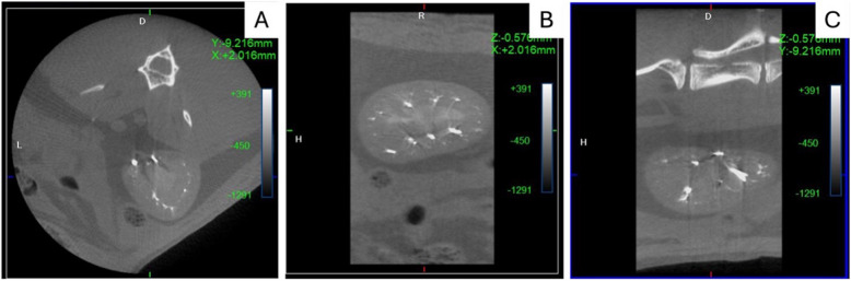 Figure 1
 Figure 2
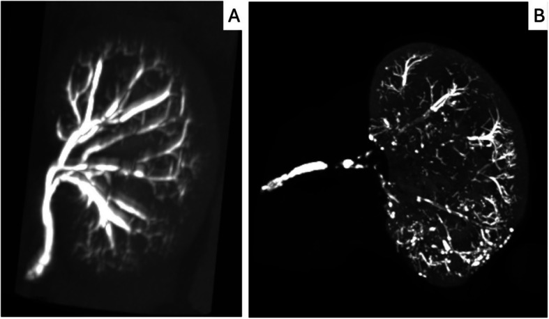 Figure 3
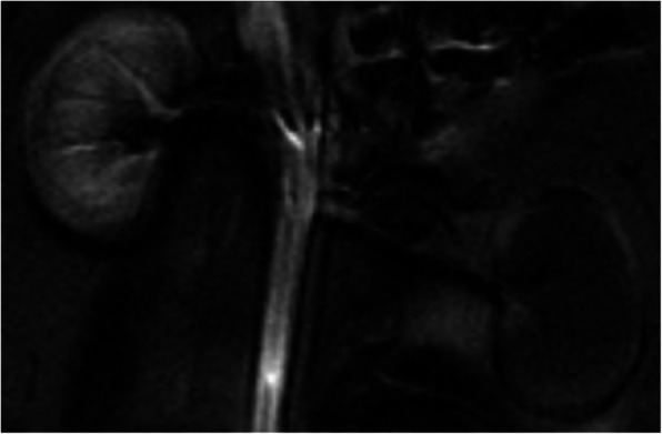 Figure 4
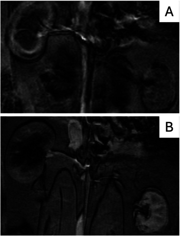 Figure 5
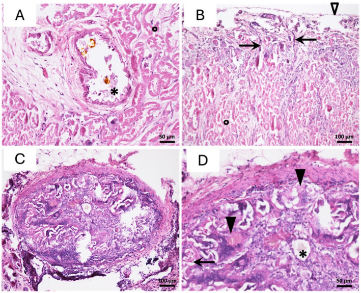 Figure 6
Peer Reviews
No public reviews on file for this paper yet. If you reviewed it on a platform where reviews are public (OpenReview, ICLR, NeurIPS, ICML), you can paste yours below so the community can read it here.
Videos
No videos yet. Explain this paper in a talk, walkthrough, or lecture? Add one.
Taxonomy
TopicsRenal and Vascular Pathologies · Renal cell carcinoma treatment · Pediatric Urology and Nephrology Studies