Cross-sectional and longitudinal quantification of total white matter perivascular space volume fraction in Dutch-type Cerebral Amyloid Angiopathy
Manon R. Schipper, Thijs W. van Harten, Arie-Tjerk Razoux-Schultz, Kanishk Kaushik, Lydiane Hirschler, Sabine Voigt, Ingeborg Rasing, Emma A. Koemans, Rosemarie van Dort, Reinier G.J. van der Zwet, Sanne E. Schriemer, Erik W. van Zwet, Jeroen van der Grond, Mark A. van Buchem

TL;DR
This study introduces a new method to measure brain perivascular spaces in a rare amyloid disease and finds increased volume in early stages.
Contribution
A novel whole-brain semi-automated method for quantifying perivascular space volume in Dutch-type Cerebral Amyloid Angiopathy.
Findings
Symptomatic D-CAA patients had significantly higher baseline PVSvf compared to younger and older controls.
Pre-symptomatic D-CAA carriers also showed increased PVSvf compared to younger controls.
No longitudinal changes in PVSvf were observed over four years between D-CAA and control groups.
Abstract
•Novel whole-brain semi-automated perivascular space segmentation-method in D-CAA.•Cross-sectional and longitudinal perivascular space volume fraction (PVSvf) in D-CAA.•Increased PVSvf in early stages of Dutch-type Cerebral Amyloid Angiopathy (D-CAA)•No difference in PVSvf-change over 4 yr follow-up between D-CAA and control subjects. Novel whole-brain semi-automated perivascular space segmentation-method in D-CAA. Cross-sectional and longitudinal perivascular space volume fraction (PVSvf) in D-CAA. Increased PVSvf in early stages of Dutch-type Cerebral Amyloid Angiopathy (D-CAA) No difference in PVSvf-change over 4 yr follow-up between D-CAA and control subjects. Enlarged perivascular spaces (PVS) in the centrum semiovale are an important marker of Cerebral Amyloid Angiopathy (CAA) and are thought to reflect brain clearance dysfunction. However, the current golden standard for…
Genes, proteins, chemicals, diseases, species, mutations and cell lines named across the full text — each resolved to its canonical identifier and authoritative record.
Click any figure to enlarge with its caption.
 Figure 1
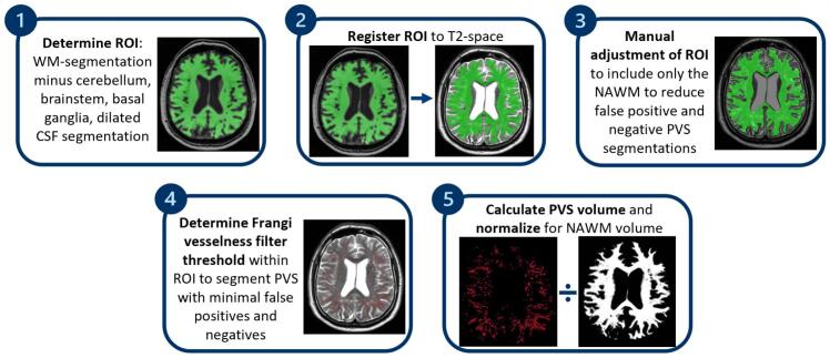 Figure 2
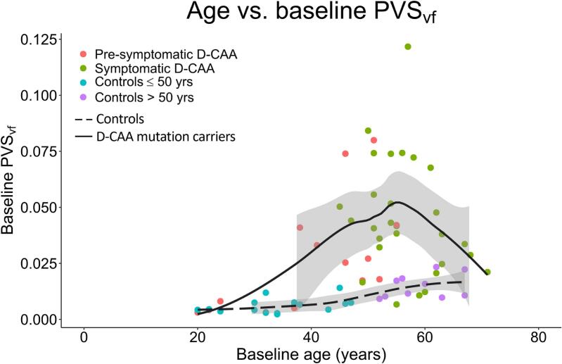 Figure 3
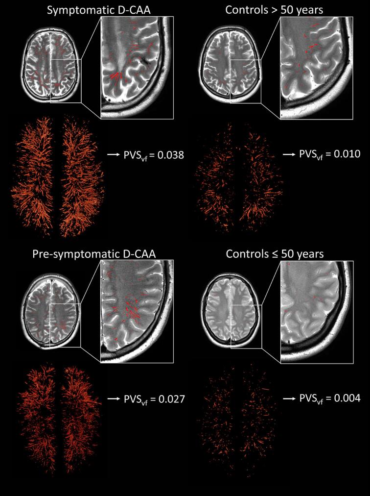 Figure 4
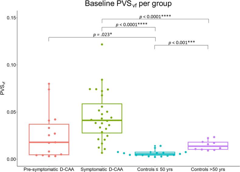 Figure 5
 Figure 6
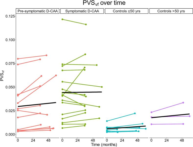 Figure 7
Peer Reviews
No public reviews on file for this paper yet. If you reviewed it on a platform where reviews are public (OpenReview, ICLR, NeurIPS, ICML), you can paste yours below so the community can read it here.
Videos
No videos yet. Explain this paper in a talk, walkthrough, or lecture? Add one.
Taxonomy
TopicsAcute Ischemic Stroke Management · Intracerebral and Subarachnoid Hemorrhage Research · Advanced Neuroimaging Techniques and Applications