How Calcifications Guide the Diagnosis: A Case of Gorlin's Cyst
Rym Kammoun, Manel Gharbi, Rawia Jaziri, Nawress Ghadhab, Imen Chaabani, Touhami Ben Alaya

TL;DR
This paper discusses how calcifications in Gorlin's cyst help diagnose and treat a benign jaw tumor.
Contribution
The study emphasizes the importance of calcification patterns in diagnosing Gorlin's cyst using imaging.
Findings
Gorlin's cyst appears as a well-limited osteolytic image with peripheral calcifications in imaging.
Recognizing these radiological features is crucial for accurate diagnosis and treatment planning.
Abstract
Calcified odontogenic epithelial cyst known as Gorlin's cyst is one of the benign odontogenic tumors of the maxillae. In imaging, the most revealing aspect is a well‐limited osteolytic image with peripheral calcifications. The aim of the study was to highlight these radiological features to establish the correct diagnosis and appropriate treatment.
Genes, proteins, chemicals, diseases, species, mutations and cell lines named across the full text — each resolved to its canonical identifier and authoritative record.
Click any figure to enlarge with its caption.
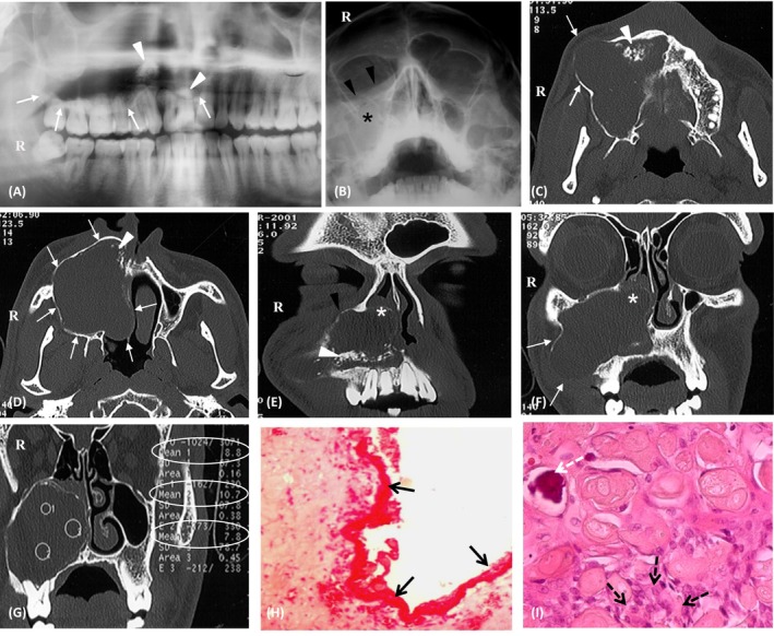 Figure 1
Peer Reviews
No public reviews on file for this paper yet. If you reviewed it on a platform where reviews are public (OpenReview, ICLR, NeurIPS, ICML), you can paste yours below so the community can read it here.
Videos
No videos yet. Explain this paper in a talk, walkthrough, or lecture? Add one.
Taxonomy
TopicsOral and Maxillofacial Pathology · Bone Tumor Diagnosis and Treatments · Hedgehog Signaling Pathway Studies