Treatment of an endodontic-periodontal lesion using peripheral blood mesenchymal stem cells (PBMSCs) and platelet-rich fibrin matrix (PRFM): A case report
Sphoorthi Anup Belludi, Sharaz Shaik, Neha Pradhan, Sreeparvathy Rema

TL;DR
This case report shows successful treatment of a complex dental lesion using a patient's own stem cells and platelet-rich fibrin, leading to significant tissue regeneration.
Contribution
The novel use of autogenous PBMSCs and PRFM for periodontal-endodontic regeneration is demonstrated in a clinical case.
Findings
Significant osseous fill was observed after nine months of treatment.
A 5 mm gain in clinical attachment level was achieved.
The combination therapy showed periodontal regenerative potential.
Abstract
In the current report, we discuss the available treatment options and present a successfully treated periodontal-endodontic lesion using autogenous peripheral blood mesenchymal stem cells (PBMSCs) and platelet-rich fibrin matrix (PRFM). A patient presented with a complaint of food impaction and bad breath. Clinically, the lower right first molar was non-vital and had a deep periodontal pocket and attachment loss. Radiographically, the distal root had an angular bone loss extending to the apex. The endodontic condition was treated with chemomechanical debridement, calcium hydroxide dressing, and obturation. Later, we reflected a full-thickness mucoperiosteal flap and thoroughly debrided the granulation tissue. We filled the defect with a gel containing PBMSCs and PRFM, prepared from the patient’s peripheral blood, and sutured the flap. After nine months, we noticed significant osseous…
Genes, proteins, chemicals, diseases, species, mutations and cell lines named across the full text — each resolved to its canonical identifier and authoritative record.
Click any figure to enlarge with its caption.
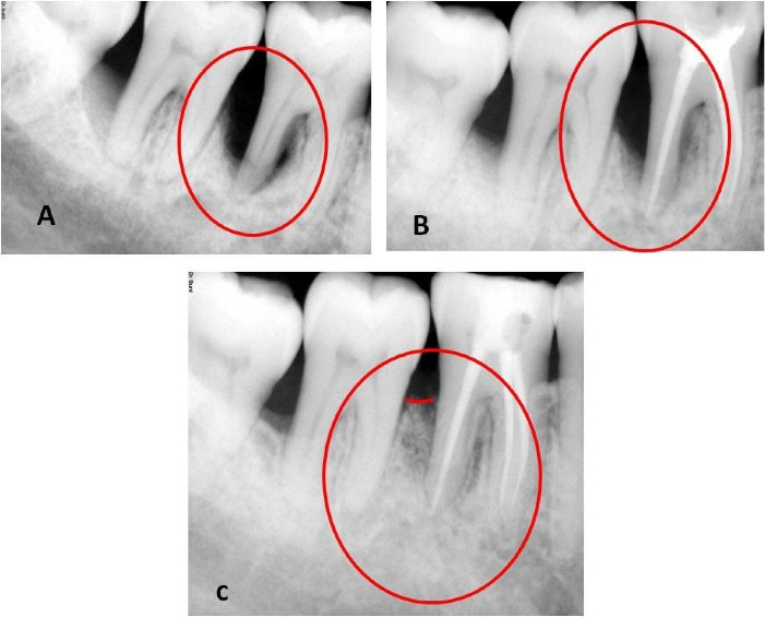 Figure 1
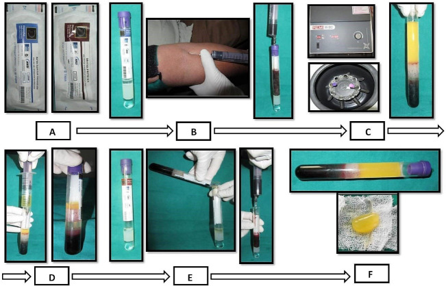 Figure 2
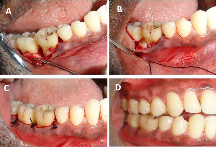 Figure 3
|
| ||||||||||||||||
|
|
|
|
| |||||||||||||
|
|
|
|
|
|
|
|
| |||||||||
| Buccal | 3 | 3 | 3 | 3 | 3 | 3 | 3 | 3 | ||||||||
| Lingual | 3 | 3 | 4 | 3 | 3 | 3 | 3 | 3 | ||||||||
| Interdental | M | D | M | D | M | D | M | D | M | D | M | D | M | D | M | D |
| 3 | 4 | 2 | 3 | 4 | 9 | 3 | 4 | 8 | 8 | 4 | 4 | 7 | 3 | 3 | 3 | |
|
|
|
| Type of radiograph | Intraoral direct digital periapical radiovisiograph |
| Radiographic equipment | RVG-Suni Medical Imaging, Apteryx Inc., Acron, Ohio, USA. |
| Technique | long cone paralleling technique |
| Exposure | 70 kVp, 7 ma for 0.2 seconds |
| The focus-to-film distance | 20 cm |
| Software for linear measurements | Image J software, Wayne Rasband, National Institute of Health, USA |
Peer Reviews
No public reviews on file for this paper yet. If you reviewed it on a platform where reviews are public (OpenReview, ICLR, NeurIPS, ICML), you can paste yours below so the community can read it here.
Videos
No videos yet. Explain this paper in a talk, walkthrough, or lecture? Add one.
Taxonomy
TopicsPeriodontal Regeneration and Treatments · Endodontics and Root Canal Treatments · Dental Trauma and Treatments
Introduction
The concomitant presence of inflammatory periodontal disease and pulpal pathosis complicates the diagnosis and treatment planning of endodontic-periodontal lesions (EPLs). Hence, EPLs usually need a multidisciplinary approach.^1,2^ The aim of treating the periodontal pocket is to regain the periodontal attachment; it may range from oral prophylaxis to regenerative procedures. Currently, much interest has been observed in autologous platelet/fibrin biologics. According to Dohan’s classification, our material of interest is pure platelet-rich fibrin/platelet-rich fibrin matrix (PRFM).^3,4^
In alignment with regenerative dentistry and the current concept of tissue engineering, stem cells can be an effective module due to their pluripotency and regenerative ability.^5^ The extraction of MSCs from bone marrow and other sources is an invasive, high-risk approach that provides a low-frequency and heterogeneous population. Peripheral blood is possibly the most straightforward source due to the accessibility and lack of donor-organ morbidity in comparison to other sources (bone marrow, umbilical cord, adipose tissue, salivary glands, periosteum, periodontium, and dental pulp) of mesenchymal stem cells (MSCs). Despite numerous studies on using other sources of stem cells in periodontal regeneration,^5^ there is a paucity of reports on the application of peripheral blood-derived mesenchymal stem cells (PBMSCs). The current case report describes a novel approach using PBMSCs + PRFM en masse to treat an EPL patient.
Case Presentation
A healthy patient (male, age = 42 years) presented with a chief complaint of discomfort and food impaction in the lower right back teeth region and bad breath for five months. Intraoral examination revealed a full constituent dentition, generalized moderate accumulation of dental biofilm, calculus, and generalized bleeding on probing, more prominent in the lower right second premolar to the third molar region (#45 to #48). Food impaction was observed between the lower right first and second molars (#46 and #47). Mild tenderness on percussion (TOP) was elicited with #46 and #47. Grade I mobility and occlusal and buccal caries were detected with #46. The clinical attachment level (CAL) and probing pocket depth (PPD) were 8 mm in the interdental region of #47 and #48 and 4 mm between #45 and #46. However, the CAL was 9 mm, and PPD was 8 mm in the interdental region of #46 and #47 due to 1 mm of gingival recession on the distobuccal root of #46 (Table 1). A long-cone paralleling technique and intraoral direct digital periapical radiovisiograph (RVG-Suni Medical Imaging, Apteryx Inc., Acron, Ohio, USA) were used for radiographic evaluation (Table 2). The radiographs revealed angular bone loss in the lower right first and second molar (#46 and #47) region, with the base of the vertical defect extending to the apex of the distal root of the lower right first molar (#46); however, there was no sign of root damage (Figure 1). Vitality test exhibited a delayed response with #46 and a positive response with #47. The patient was devoid of complicating factors such as diabetes and smoking, which could have affected the healing and treatment outcome. Based on the presentations and common classification by Simon et al.,^6^ the lesion was classified as primary periodontal lesions with secondary endodontic involvement and grade 3 endo-periodontal lesion in periodontitis patients as per the 2017 World Workshop Classification.^7^
Treatment
Regarding the clinical and radiographic findings, the provisional prognosis was considered fair. The treatment protocol was planned in 4 phases^8^: (1) preliminary management, (2) endodontic management, 3: PBMSCs + PRFM gel preparation and surgical procedure, and (4) follow-up management.
Preliminary management and patient consent
We performed oral prophylaxis, educated the patient about the importance of oral hygiene, and demonstrated the use of dental floss and chlorhexidine mouthwash. Since the patient did not have acute symptoms, we did not prescribe oral medications. We explained the current condition of the lower right first molar (#46) and our proposal to use the novel treatment procedure. The patient voluntarily gave consent for the procedure. Written informed consent was obtained from the patient for enrollment into the treatment protocol and for publishing the acquired data. Ethical clearance for the treatment was received from the Institutional Ethics Committee (Blinded for review).
Endodontic management
Root canal treatment incorporated chemomechanical debridement under local anesthesia and isolation using rotary endodontic instruments (ProTaper Next, Dentsply Sirona) and filling of the root canals with calcium hydroxide paste (ApexCal® Ivoclar Vivadent) using a lentulo spiral. The root canals were obturated by lateral condensation of gutta-percha (ProTaper Next Gutta Percha endodontic points, Dentsply, Maillefer) and a calcium hydroxide-based sealer (Apexit Plus Root Canal Sealer, Ivoclar Vivadent Inc.). A permanent composite resin restoration was placed to seal off the access cavity.
PBMSCs + PRFM gel preparation9
PBMSCs and PRFM were obtained from the peripheral blood by Merisis Supercell concentrate, DiponEd BioIntelligence© and Merisis PRFM kit, Merisis Biological Devices, and DiponEd BioIntelligence©, respectively, by following manufacturer kit insert instructions (Figure 2).^9^
Surgical procedure
After anesthetizing with 2% lignocaine containing 1:80,000 adrenaline (Lignox A 2%, Indoco Remedies LTD., India) using the inferior alveolar nerve block. We placed sulcular incisions on both buccal and lingual sides and reflected a full-thickness mucoperiosteal flap extending from the mesial of the lower right second premolar (#45) to the mesial line angle of the lower right third molar (#48). Thorough debridement of granulation tissue in the intrabony defect (3-wall defect between #46 and #47 and #47 and #48 regions), scaling, and root planing of remnant calculus on the root surfaces was performed (Figure 3). After pre-suturing of the interdental papillae in the #46, #47, and later in #47 and #48 regions, the freshly prepared PBMSCs + PRFM was extracted en masse from the glass vial with the help of tweezers and filled into the intrabony defect as a sole material. The flaps were approximated by interrupted sutures using 4-0 silk material (Figure 3). No periodontal dressing was applied. Antibiotics (Amoxycillin 500 mg every 8 hours for 7 days) and analgesics (Ibuprofen 400 mg every 4 hours) as required for pain management were prescribed. 0.2% chlorhexidine rinses every 12 hours for 14 days were advised. Postoperative instructions were given, an ultra-soft toothbrush was prescribed, and the patient was advised to refrain from disturbing the surgical site for the next two days.
Outcome and follow-up
The patient was followed for one month after endodontic treatment with no evident changes in the clinical parameters; hence, we proceeded to surgical therapy. We also conducted post-surgical follow-up after a week, one month, 3 months, and 9 months. During the first follow-up appointment, the patient had no complaints of pain and discomfort and no tenderness on percussion. We reinforced oral prophylactic instructions. The sutures were removed ten days after surgery. During the one-month and three-month appointments, the patient displayed good oral hygiene. The surgical area displayed complete healing, no tenderness on percussion, and no tooth mobility. Radiographic evaluation three months after surgery displayed a partial bone fill (Figure 1). During the 9-month follow-up appointment, the periodontal lesion had significantly improved clinically and radiographically (Figures 1 and 3). A significant reduction in the periodontal probing depth and gain in the clinical attachment level was noted (Table 1). At any of these intervals, there was no attempt at subgingival instrumentation.
**
**
**
Discussion
A combined EPL does not respond with root canal treatment alone. The patient’s 4-week post-root canal treatment follow-up evaluation did not indicate any distinct improvement in the clinical parameters. Considering CAL deeper than the critical probing depth of 5.4 mm in our case, surgical intervention with regenerative therapy was deemed beneficial.^10^ In the current case, after initial biomechanical debridement, the canals were filled with calcium hydroxide endodontic dressing for a week, followed by obturation with gutta-percha and a calcium hydroxide-based sealer. A report suggests that pulpal infection tends to stimulate epithelial growth apically adjacent to the stripped dentinal surface, especially in the presence of periodontal infection.^10,11^ Open dentinal tubules, accessory canals, and apical foramen are the three possible transmission channels for contamination.^12^
In the present case, as the vitality test showed a delayed response, endodontic therapy was carried out to eliminate the partially necrotic pulp and prevent remnant nidus of infection and future necrosis and infection following periodontal regenerative surgery. Therefore, root canal treatment was completed before periodontal regenerative therapy. Furthermore, calcium hydroxide dressing in the root canal could promote periodontal healing in such cases.^10^ The overall success of an EPL usually depends on the efficiency of periodontal therapy.
Numerous biomaterials are currently available for periodontal regeneration. A study reported that a combination of PRP and stem cells resulted in better periodontal tissue regeneration than stem cell therapy alone^13^; furthermore, using PRFM instead of PRP could produce better results.^14,15^ PRFM enhances osteoblast differentiation and stimulates periodontal soft tissue regeneration.^16^ The influence of PRFM on periodontal regeneration can be accredited to transforming growth factor β, platelet-derived growth factor, insulin-like growth factor, and basic fibroblast growth factor.^15,17^ Apart from the healing potential of PRFM, it was shown that PRFM produced significantly higher mesenchymal stem cell proliferation.^17^ There are studies on the application of stem cells from various sources, more so on bone marrow mesenchymal stem cells;^5^ however, little is known on tissue regeneration by PBMSCs. PBMSCs have equivalent pluripotency and osteogenic ability to mesenchymal stem cells derived from bone marrow and umbilical cord.^18,19^ The stem cell-based periodontal regeneration could be mediated by monocyte chemoattractant protein-1, chemokine stromal cell-derived factor-1, chemokine stromal cell-derived factor-1, and exosomes cell-free strategy.^20^ Combining PRFM with PBMSCs would be more salutary than PRP as there is a more sustained release of growth factors by PRFM in comparison to PRP.^21,22^ Our approach of using PBMSCs + PRFM en masse in periodontal regeneration of EPL is novel as there are no similar reports in the current scientific literature. However, few studies suggest promising results for this combination as a regenerative material.^9,13^
The success rate of conventional modalities in the treatment of EPL combined lesions was reported to be very low (27‒37%) compared to a tooth survival rate of 92.31% at 5 years after a periodontal regenerative procedure.^23,24^Studies suggest that treatment outcomes seen at a first-year follow-up appointment tend to last for a long time.^25^ Our 9-month follow-up revealed an excellent treatment outcome. Uniformity in pre- and postoperative radiography and analysis were used to minimize the errors (Table 2). No bone graft or radiopaque material was used, which might mimic radiopaque bone fill in postoperative radiographs. Hence, the postoperative radiopacity can be considered a true osseous fill.
Conclusion
A substantial osseous fill in the present study can be attributed to a definitive root canal treatment, periodontal debridement, and application of PRFM + PBMSCs. This protocol can be an easy, effective, and promising regenerative option. However, in vitro validity tests, histomorphometry studies, and large-scale randomized control trials are required to endorse the regenerative efficacy of this protocol.
Competing Interests
The authors do not have any financial interest in the companies whose materials are included in this article and have no conflicts of interest.
Consent for Publication
Not applicable.
Data Availability Statement
Not applicable.
Ethical Approval
We explained the current condition and our proposal for using the novel treatment procedure. The patient voluntarily gave consent for the procedure. Written informed consent was obtained from the patient for enrollment into the treatment protocol and for publishing the acquired data. Ethical clearance for the treatment was received from the Institutional Ethics Committee (IEC/Nov-2021/8).
The reference list from the paper itself. Each links out to its DOI / PubMed record.
- 1Tayal A Ghosh S Adhikari HD Ghosh A Management of an endo-perio lesion: a multidisciplinary approach IP Indian J Conserv Endod 202163171510.18231/j.ijce.2021.037 · doi ↗
- 2Al Attas MA Edrees HY Sammani AM Madarati AA Multidisciplinary management of concomitant pulpal and periodontal lesion: a case report J Taibah Univ Med Sci 20171254556010.1016/j.jtumed.2017.05.01031435278 PMC 6694962 · doi ↗ · pubmed ↗
- 3Dohan Ehrenfest DM Andia I Zumstein MA Zhang CQ Pinto NR Bielecki T Classification of platelet concentrates (platelet-rich plasma-PRP, platelet-rich fibrin-PRF) for topical and infiltrative use in orthopedic and sports medicine: current consensus, clinical implications and perspectives Muscles Ligaments Tendons J 2014413924932440 PMC 4049647 · pubmed ↗
- 4Simon BI Gupta P Tajbakhsh S Quantitative evaluation of extraction socket healing following the use of autologous platelet-rich fibrin matrix in humans Int J Periodontics Restorative Dent 20113132859521556385 · pubmed ↗
- 5Bassir SH Wisitrasameewong W Raanan J Ghaffarigarakani S Chung J Freire M Potential for stem cell-based periodontal therapy J Cell Physiol 20162311506110.1002/jcp.2506726058394 PMC 4627700 · doi ↗ · pubmed ↗
- 6Simon JH Glick DH Frank AL The relationship of endodontic-periodontic lesions J Periodontol 1972434202810.1902/jop.1972.43.4.2024505605 · doi ↗ · pubmed ↗
- 7Papapanou PN Sanz M Buduneli N Dietrich T Feres M Fine DH Periodontitis: consensus report of workgroup 2 of the 2017 World Workshop on the Classification of Periodontal and Peri-Implant Diseases and Conditions J Periodontol 201889 Suppl 1S 1738210.1002/jper.17-072129926951 · doi ↗ · pubmed ↗
- 8Oh SL Fouad AF Park SH Treatment strategy for guided tissue regeneration in combined endodontic-periodontal lesions: case report and review J Endod 200935101331610.1016/j.joen.2009.06.00419801225 · doi ↗ · pubmed ↗