Surgical Approach to Bilateral Impacted and Inverted Mesiodentes in a Nonsyndromic Pediatric Patient: A Case Report and Brief Literature Review
Nhung T Nguyen, Quang V Dang, Vinh Q Dang

TL;DR
This case report describes the surgical removal of two unusual extra teeth in a child's upper jaw, leading to a successful outcome.
Contribution
The paper presents a rare case of bilateral inverted mesiodentes in a nonsyndromic pediatric patient and advocates for early surgical intervention.
Findings
Bilateral impacted and inverted mesiodentes were successfully managed through surgical extraction and orthodontic treatment.
Timely surgical intervention led to satisfactory functional and aesthetic outcomes for the patient.
Periodic clinical and radiographic evaluations are emphasized for early detection and management of supernumerary teeth.
Abstract
Supernumerary teeth (ST) are a form of abnormal dental development and may not always present with symptoms. This case report discusses an eight-year-old child who presented with spacing in the maxillary anterior region. Radiographic examination revealed bilateral impacted and inverted mesiodentes, which were successfully managed through surgical extraction and orthodontic treatment. The postoperative course was uneventful, resulting in satisfactory outcomes for both the patient and his parents. We emphasize the importance of periodic check-ups during the early stages of tooth exfoliation, including proactive clinical and radiographic evaluations for the early diagnosis and management of multiple ST. Along with a review of the literature, this report suggests that a timely surgical approach should be indicated to effectively address the patient’s condition, ensuring optimal outcomes…
Genes, proteins, chemicals, diseases, species, mutations and cell lines named across the full text — each resolved to its canonical identifier and authoritative record.
Click any figure to enlarge with its caption.
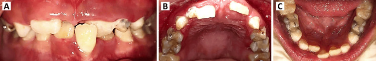 Figure 1
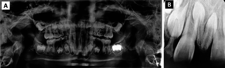 Figure 2
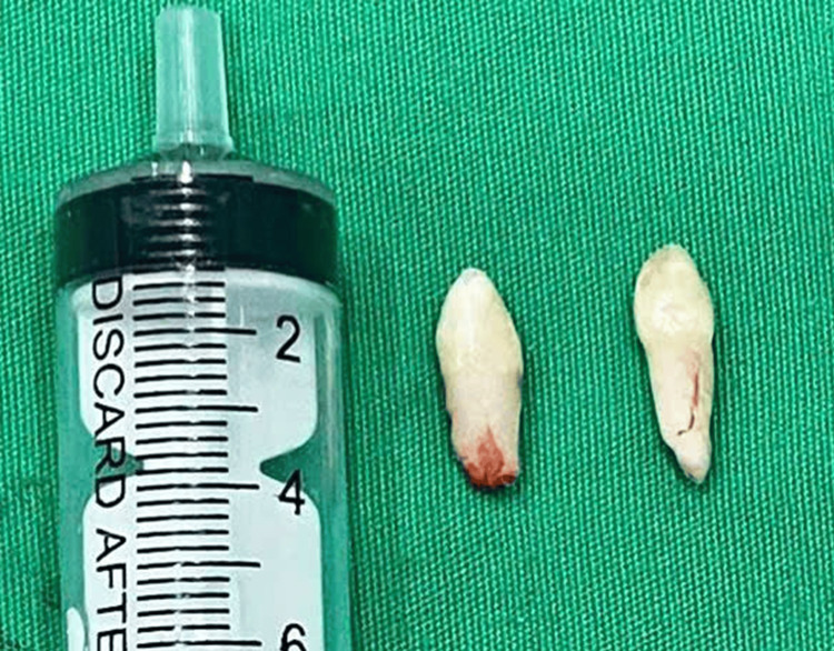 Figure 3
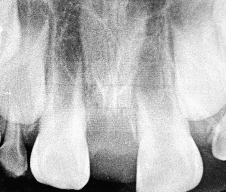 Figure 4
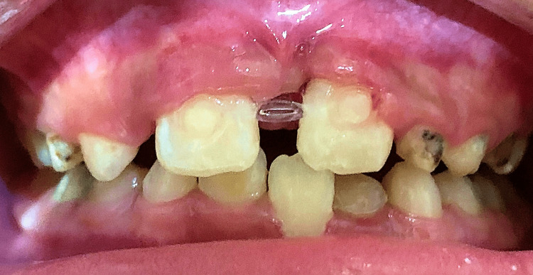 Figure 5
| Case | Study | Year | Country | Age | Sex | Chief complaint |
| 1 |
Dinkar et al. [ | 2007 | India | 14 years | Female | Painful palatal swelling |
| 2 |
Canoglua et al. [ | 2009 | Türkiye | 8 years | Male | Maxillary anterior crowding |
| 3 |
Byatnal et al. [ | 2013 | India | 13 years | Male | Maxillary anterior swelling |
| 4 |
Krishnappa et al. [ | 2014 | India | 16 years | Female | Maxillary anterior misalignment |
| 5 |
Desai et al. [ | 2014 | India | 12 years | Male | Proclined maxillary anterior teeth |
| 6 | 7 years | Male | Maxillary anterior teeth uneruption | |||
| 7 | 25 years | Female | Discolored tooth on right maxilla | |||
| 8 |
Viswanathan and Pai [ | 2015 | India | 13 years | Male | Palatal swelling and pain during swallowing |
| 9 |
Al-Sehaibany et al. [ | 2016 | Saudi Arabia | 8.5 years | Male | Delayed eruption of maxillary anterior teeth |
| 10 |
Sharifi et al. [ | 2021 | Iran | 9 years | Male | Routine dental checkup |
| 11 |
Rajaram Mohan et al. [ | 2022 | India | 21 years | Male | Maxillary anterior absence |
| 12 |
Koyama et al. [ | 2023 | Japan | 9 years | Male | Malocclusion |
| 13 | This case | 2025 | Vietnam | 8 years | Male | Maxillary anterior spacing |
| Case | Study | Year | Radiographic type | Clinical findings | Treatment | Follow-up |
| 1 |
Dinkar et al. [ | 2007 | Pano, Occl | Dentigerous cyst | SE | 6 months |
| 2 |
Canoglua et al. [ | 2009 | Pano, Occl, PA, LC | Crowding | SE, OT | 24 months |
| 3 |
Byatnal et al. [ | 2013 | CT | Dentigerous cyst | SE | N/A |
| 4 |
Krishnappa et al. [ | 2014 | Pano, Occl, PA, LC | Crowding | SE, OT | N/A |
| 5 |
Desai et al. [ | 2014 | Pano | Proclined anterior teeth | N/A | N/A |
| 6 | Occl | Proclined anterior teeth | N/A | N/A | ||
| 7 | Occl | Proclined anterior teeth | N/A | N/A | ||
| 8 |
Viswanathan and Pai [ | 2015 | Occl | Swelling on palatal vault | SE | 12 months |
| 9 |
Al-Sehaibany et al. [ | 2016 | Pano, Occl, CBCT | Delayed central incisor eruption | SE, OT | 12 months |
| 10 |
Sharifi et al. [ | 2021 | Pano, CBCT | Proximity to anterior nasal spine and tooth roots | SE | N/A |
| 11 |
Rajaram Mohan et al. [ | 2022 | Pano, CBCT | Promixity of anterior nasal spine | N/A | N/A |
| 12 |
Koyama et al. [ | 2023 | Pano, CT | Promixity of tooth root | SE/mixed reality | 12 months |
| 13 | This case | 2025 | Pano, PA | Diastema | SE, OT | 6 months |
Peer Reviews
No public reviews on file for this paper yet. If you reviewed it on a platform where reviews are public (OpenReview, ICLR, NeurIPS, ICML), you can paste yours below so the community can read it here.
Videos
No videos yet. Explain this paper in a talk, walkthrough, or lecture? Add one.
Taxonomy
TopicsOral and Maxillofacial Pathology · Cleft Lip and Palate Research · dental development and anomalies
Introduction
Supernumerary teeth (ST), also referred to as hyperdontia, is a developmental anomaly characterized by an excessive number of teeth in addition to normality. ST can be single or multiple, as well as symmetric or asymmetric in any region of the dental arch [1]. The most common type of ST is mesiodens, which emerges between the maxillary central incisors [2]. Several theories have been proposed to explain the development of ST, including atavism, dichotomy, and hyperactivity of the dental lamina [3]. In addition, various oral disorders and hereditary syndromes have been associated with its occurrence [4]. However, the precise pathological mechanisms of ST remain poorly understood [3].
ST-related complications may include dental impaction, ectopic eruption, delayed eruption, malocclusion, root dilaceration, and cyst formation [1]. ST may be retained if they do not cause any adverse effects or may be considered as replacement teeth if they fulfill a functional and aesthetic role in the absence of the relevant permanent teeth [5]. However, surgical extraction is frequently indicated to address these complications. Herein, we present a nonsyndromic case involving bilateral impacted and inverted mesiodentes, which was successfully treated with surgical extraction followed by orthodontic treatment. This report aims to contribute a typical case of bilateral impacted and inverted mesiodentes, accompanied by a brief literature review, emphasizing the importance of early treatment to address both the pathological and cosmetic aspects of the patient’s condition.
Case presentation
An eight-year-old Vietnamese boy accompanied by his mother to visit the Department of Odonto-Stomatology with a chief complaint of spacing in the maxillary anterior region. His medical and dental history revealed no associated systemic diseases or allergies. Intraoral examination displayed a very large diastema (approximately 9 mm) between permanent maxillary central incisors, a crossbite between the permanent maxillary left central incisor and the permanent mandibular left central incisor, as well as the presence of dental plaque, tartar, and caries (Figure 1).
Intraoral photographs taken at the initial examination. (A) Frontal view. (B) Maxillary occlusal view. (C) Mandibular occlusal view
Radiographic examination, including panoramic and periapical radiographs, revealed the presence of two impacted and inverted ST between the permanent maxillary central incisors, which resulted in a diastema. Only one of these teeth exhibited nearly complete root formation (Figure 2). The proposed comprehensive treatment plan included surgical extraction, followed by orthodontic intervention.
Preoperative radiographs revealing bilateral impacted and inverted mesiodentes. (A) Panoramic radiograph. (B) Periapical radiograph
During the preoperative treatment, the child underwent scaling with an ultrasonic scaler. The surgical procedure was performed under local anesthesia and aseptic conditions. A three-sided flap was created using a periosteal elevator, a scalpel handle #3, and a scalpel blade #15. A low-speed handpiece with a No. 702 bur was utilized under abundant saline irrigation. The mesiodentes were removed by an elevator and forceps with the entire crowns and roots (Figure 3). The extraction socket was gently probed by a curette and flushed with saline to remove any surgical debris. The final step is the closure of the socket, where the flap was repositioned with a 3-0 nylon suture.
Bilateral mesiodentes extracted with the entire crowns and rootsSyringe placed for scale
The patient returned for suture removal, and a periapical radiograph was taken after two weeks, revealing good healing (Figure 4). The patient was then referred for orthodontic treatment to address the crossbite and diastema issues and continued to receive appropriate monitoring of the results over a six-month period (Figure 5).
Periapical radiograph taken two weeks after surgery
Intraoral photograph taken within one week of orthodontic intervention
Discussion
The prevalence of ST ranges from 0.3% to 0.8% in deciduous dentition and from 1.5% to 3.5% in permanent dentition [1]. ST are more commonly found in the maxilla than the mandible and more frequently in men than women [4]. Among studies of the Asian population, ST are typically found as a single tooth in children with mixed dentition. Mesiodens, inverted direction, impacted status, and conical shape are the predominantly reported characteristics [2,6]. The literature review includes 13 reported cases of bilateral impacted and inverted mesiodentes in English to date, including the current case [7-16]. All of these cases were observed in Asian countries, with a male-to-female ratio of 10:3. More than half of the cases were in children aged 6-12 years, while the remaining cases involved older individuals, with the maximum age of 25. Most patients typically seek hospital or clinic care when complications arise (Table 1). Unlike other cases in the literature review, the case reported by Sharifi et al. was exceptional, as it involved the incidental recognition of bilateral impacted and inverted mesiodens during a routine dental check-up [14].
Radiographic examination is a crucial and indispensable procedure for identifying ST regardless of the radiographic modality [17]. Table 2 presents the types of radiographic tools utilized in case reports from the literature review. While panoramic radiographs are commonly employed, they are not considered the most appropriate tool for identifying ST, and additional radiographs may be required for a precise diagnosis [18]. It is noteworthy that cone-beam computed tomography (CBCT) has recently been highlighted as the most effective application for ST diagnosis, offering accurate information on the number, shape, location, and proximity of vital neighboring anatomical structures [19]. In our department, although CBCT was not employed to identify the bilateral impacted and inverted mesiodentes due to the unavailability of advanced technical facilities, the combination of panoramic radiographs and periapical radiographs represents a cost-effective and practical diagnostic option for patient care.
Among the ST-related complications observed in cases of bilateral impacted and inverted mesiodentes, crowding, diastema, delayed eruption, and proclination of adjacent permanent teeth were common, whereas two patients experienced significant issues with dentigerous cysts. Although the optimum timing for impacted ST extraction has been debated, some advocate for early diagnosis and intervention of impacted mesiodens to minimize future complications and the need for orthodontic treatment [19,20]. In terms of treatment, the unusual number, direction, and proximity to vital neighboring anatomical structures in these cases could increase the potential for exacerbated effects and necessitate specialized care if timely intervention is not provided. Nine out of 11 authors reported surgical extraction as the intervention method, while only two did not mention the type of procedure conducted in their patients. In the present case, the patient is in the ugly duckling stage, and a combination of surgical extraction and orthodontic treatment is considered the most appropriate option. We believe the surgical approach should be prioritized in cases of this condition due to the heightened risk involved.
Although the postsurgical period is also crucial to managing multiple ST, only about half of the reported cases included a monitoring duration of 6 to 24 months. In this case, the patient has been observed for six months, with a focus on bone healing progress, closure of the midline diastema, and proper eruption of remaining teeth, particularly the maxillary canines. The patient has not experienced any discomfort, and both he and his parents have expressed satisfaction with the positive outcome.
Despite the lack of advanced radiographic techniques, the current patient was successfully treated with a timely surgical approach and minimal orthodontic intervention, effectively avoiding significant ST-associated complications reported in other cases in the literature review. Nonetheless, we underscore that the utilization of CBCT should be strongly considered for comprehensive preoperative assessment in future research and clinical practice improvements, given its crucial role in evaluating the proximity to vital structures and aiding in surgical orientation. Furthermore, the literature review highlights that bilateral impacted and inverted mesiodentes may predominantly be observed only in Asian populations. However, this may not fully represent the global distribution of this condition due to the exclusion of case reports published in non-English languages or those with restricted access to full texts, which presents another limitation of our study.
Conclusions
Most cases of bilateral impacted and inverted mesiodentes are not promptly identified until complications arise, including the present case. Therefore, surgical extraction should be regarded as the optimal treatment for addressing both aesthetic and pathological concerns in those individuals. Periodic check-ups, including comprehensive clinical and radiographic examinations, are essential for the early diagnosis and management of ST. Future research and clinical practitioners should recognize CBCT as a preferred radiographic modality for ST management, particularly in the preoperative evaluation of proximity to vital neighboring structures and surgical orientation.
The reference list from the paper itself. Each links out to its DOI / PubMed record.
- 1Prevalence, etiology, diagnosis, treatment and complications of supernumerary teeth J Clin Exp Dent Ata-Ali F Ata-Ali J Peñarrocha-Oltra D Peñarrocha-Diago M 086201410.4317/jced.51499 PMC 428291125593666 · doi ↗ · pubmed ↗
- 2Analysis of the distribution of supernumerary teeth and the characteristics of mesiodens in Bengbu, China: a retrospective study Oral Radiol Zhao L Liu S Zhang R Yang R Zhang K Xie X 2182233720213219866310.1007/s 11282-020-00432-3 · doi ↗ · pubmed ↗
- 3Aetiology of supernumerary teeth: a literature review Eur Arch Paediatr Dent Anthonappa RP King NM Rabie AB 2792881420132406848910.1007/s 40368-013-0082-z · doi ↗ · pubmed ↗
- 4Supernumerary teeth: review of literature and decision support system Indian J Dent Res Amarlal D Muthu MS 1171222420132385224410.4103/0970-9290.114911 · doi ↗ · pubmed ↗
- 5Tooth number abnormality: from bench to bedside Int J Oral Sci Zhang H Gong X Xu X Wang X Sun Y 51520233660440810.1038/s 41368-022-00208-x PMC 9816303 · doi ↗ · pubmed ↗
- 6Prevalence of supernumerary teeth and its associated complications among school-going children between the ages of 6 and 15 years of Jamshedpur, Jharkhand, India Int J Clin Pediatr Dent Singh AK Soni S Jaiswal D Pani P Sidhartha R Nishant Nishant 5045081520223686572110.5005/jp-journals-10005-2442 PMC 9973100 · doi ↗ · pubmed ↗
- 7Dentigerous cyst associated with multiple mesiodens: a case report J Indian Soc Pedod Prev Dent Dinkar AD Dawasaz AA Shenoy S 56592520071745697210.4103/0970-4388.31994 · doi ↗ · pubmed ↗
- 8Double inverted mesiodentes: report of an unusual case Eur J Dent Canoglua E Erb N Cehrelic ZC 2192233200919756197 PMC 2741194 · pubmed ↗