A Rare Case of Atypical Choroid Plexus Papilloma in an Adult Male Patient: A Case Report
Abebe Melis Nisiro, Teketel Tadesse Geremew

TL;DR
A 22-year-old man with an unusual brain tumor in the lateral ventricle underwent surgery, and the tumor was identified as a WHO Grade II atypical choroid plexus papilloma.
Contribution
This case report highlights a rare instance of atypical choroid plexus papilloma in an adult male patient.
Findings
The patient presented with abnormal body movement and headache.
Surgical resection revealed a WHO Grade II atypical choroid plexus papilloma.
The lesion was located in the lateral ventricle and successfully resected.
Abstract
Choroid plexus tumors (CPTs) are rare neoplasms. Patient presentation varies depending on the location of the lesions. Gross total resection of primary lesions remains the gold standard for surgical treatment of CPTs. Here, we present the case of a 22-year-old male patient with 2-day history of abnormal body movement and headache who was found to have an enhancing mass of the lateral ventricle. The patient underwent craniotomy for gross-total resection of the lesion, with final histopathology demonstrating WHO Grade II aCPP.
Genes, proteins, chemicals, diseases, species, mutations and cell lines named across the full text — each resolved to its canonical identifier and authoritative record.
Click any figure to enlarge with its caption.
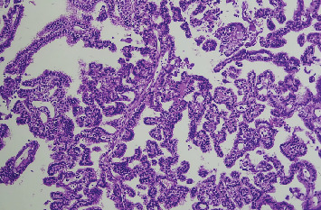 Figure 1
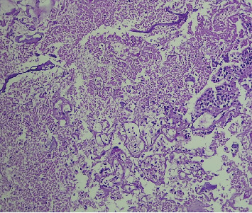 Figure 2
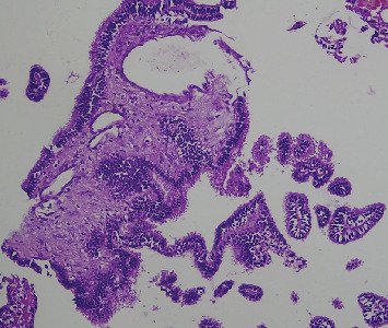 Figure 3
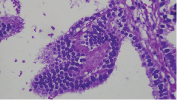 Figure 4
Peer Reviews
No public reviews on file for this paper yet. If you reviewed it on a platform where reviews are public (OpenReview, ICLR, NeurIPS, ICML), you can paste yours below so the community can read it here.
Videos
No videos yet. Explain this paper in a talk, walkthrough, or lecture? Add one.
Taxonomy
TopicsGlioma Diagnosis and Treatment · Meningioma and schwannoma management · Ocular Oncology and Treatments
1. Introduction
Choroid plexus tumors (CPTs) are papillary neoplasms derived from the choroid plexus epithelium and are typically located in the ventricles [1]. Choroid plexus papillomas (CPPs) are usually found in the lateral ventricle in children, and they are usually found in the fourth ventricle in adults [2]. Seventy-four percent of cases were found in the first 10 years of life, and 45% in the first year of life [2]. Though they only make up 0.3%–0.8% of total brain tumors, these tumors are more common in younger people, making up 2%–4% of brain tumors in children under the age of 15 and 10%–20% of those in infants under the age of 1 year [3]. CPTs are classified into three types by the World Health Organization (WHO): choroid plexus carcinoma (CPC) (malignant; WHO Grade III), CPP (WHO Grade II; with intermediate features), and benign and more differentiated CPP (WHO Grade I) [1, 4]. Atypical choroid plexus papilloma (aCPP) is defined as a CPP that has increased mitotic activity; however, it does not fulfill the criteria for malignancy (high cellularity, brisk mitotic activity, nuclear pleomorphism, blurring of the papillary growth pattern, necrosis, and diffuse brain invasion) of CPC [1, 4]. Tumors of the lateral ventricle can cause specific deficits including hemiparesis, papilledema that results in blindness, convulsions, and mental abnormalities [5, 6]. MRIs of the brain and spine (with and without gadolinium) are used to evaluate the extent of the disease in patients diagnosed with aCPP [7]. In all CPTs, complete surgical tumor resection is considered the mainstay of treatment as well as an important prognostic factor [1, 7, 8].
2. Case
A 22-year-old male patient presented with abnormal body movements of 2 days duration that occur frequently in a day and night; each episode lasts for 5–10 min with a postictal sleep state. Finally, the patient developed a loss of consciousness for 20 min, with associated high-grade persistent fever and shortness of breath. Otherwise, he has no history of trauma to the head or other sites.
On physical examination on general appearance, the patient looked acutely sick and in distress, having tachypnea, tachycardia, and febrile. On examination, there is clear media, pupils are slightly reactive, lens is transparent, fundus shows pink disc, and fine crepitation is noted over lower chest with GCS: 14/15.
For the clinical impression of an intracranial space-occupying lesion+aspiration pneumonia+status epilepticus, he was investigated with laboratory tests.
On complete blood count WBC: leukocytosis with neutrophil predominance, Hgb and platelet- normal. ESR-increased, RBS -normal, renal and liver function test- normal, serum electrolytes- normal, HBV and HCV-negative and PICT-nonreactive.
On CSF analysis, it was unremarkable. No gram stain or AFB stain showed bacilli.
Brain MRI scan showed that there is a 3.3 × 3 cm lobulated frond-like T1 isointense and T2 isointense intraventricular choroid plexus of the left ventricle with homogeneous enhancement. There is dilation of both lateral ventricles with third and fourth ventricles; the brain parenchyma is normal in signal intensity, with no evidence of mass, hematoma, or shift of midline structures, with the conclusion of the left lateral ventricle choroid plexus lobulated frond-like T1 isointense and T2 isointense with homogeneous enhancement and communicating hydrocephalus suggestive of CPP.
For this, he underwent craniotomy and gross total resection. In addition, he was put on broad-spectrum antibiotics and antiepileptic medications.
The pathology unit received specimens in 10% neutral buffered formalin.
On gross morphologic examination, there were multiple gray-white to gray-brown tissue fragments measuring 2 cm in aggregates.
Microscopic examination shows tissue composed of branching finger-like papillary architecture with fibrovascular core lined by single to pseudostratified cuboidal to columnar epithelium exhibiting moderate pleomorphism. There are two mitotic figures/10 hpf, a geographic area of necrosis, frequent psammomatoid calcification, and a focal area of blurring of papillary architecture that was evident (Figures 1, 2, 3, and 4). S100 and CK were done, and they were positive.
Subsequently, follow-up CT and MRI scans were done, which show no residual mass left, and he is on follow-up schedule every 3 months with a good clinical condition.
3. Discussion
CPTs are a diverse group of neoplasms that range from well-differentiated papillomas (WHO Grade I) to very aggressive CPCs (WHO Grade III), with rare intermediate forms known as “aCPP” whose biologic behavior remains unknown [6, 9]. aCPP are rare intermediate types of CPT, characterized as a CPP with elevated mitotic activity but does not meet the criteria for CPC [1]. aCPP was first described in 1990 [10]. In 2007, the WHO added it to the central nervous system tumor classification and classified it as WHO Class II [10–12]. It most commonly affects youngsters, with the lateral ventricle accounting for 83%, followed by the third ventricle (13%), and the fourth ventricle (3%) (Table 1) [10, 13]. Unlike the typical age, our patient is an adult. CPP's clinical presentation may include signs and symptoms of obstructive hydrocephalus, vertigo, diplopia, lateral gaze palsies, and visual field abnormalities, but vary depending on the location [14, 15]. CPT of the lateral ventricle presentation include convulsions, mental abnormalities, papilledema leading to loss of eyesight, and localized deficits including hemiparesis [16]. Symptoms of this tumor in the fourth ventricle include headache, ataxia, nystagmus, cerebellar signs, dizziness, loss of vision, vomiting, and diplopia [17]. On CT and MRI scans, CPPs usually present as isodense or hyperdense, T1-isointense, T2-hyperintense, irregularly contrast-enhancing, well-delineated masses within the ventricles, but irregular tumor margins and disseminated disease may occur [2, 18]. MRI scans cannot distinguish it from the other two choroid plexus epithelial tumors, and the diagnosis is based on histology [10]. Intraoperative observations in rare aCPPs demonstrate a highly vascular tumor with a propensity to bleed, but this feature is also observed in CPPs [7].
Currently, histological diagnostic criteria recommended by the WHO is that mitotic count in CPP > 2 mitoses per 10 randomly selected high-power fields (with one high-power field corresponding to 0.23 mm^2^); besides, one or two of the following four features may also be present: increased cellularity, nuclear pleomorphism, blurring of the papillary pattern (solid growth), and areas of necrosis; however, these features are not required for a diagnosis of aCPP [10, 19]. Based on this criteria our case mitosis of 2/10 hpf, area of necrosis and solid sheet growth pattern so it is graded as aCPP. According to the morphological diagnostic criteria above, it can be distinguished from CPP and CPC [10].
Gross total resection of primary lesions and associated implants remains the gold standard for surgical treatment of CPP [1, 13, 20]. Our patient was managed with GTR. There are no prophylactic treatments for preventing CPP metastasis [13]. Histological appearance may not always indicate biologic behavior [11]. aCPP had a 5-year survival rate of 89%, which was between CPP and CPC [10]. Metastatic disease is even less common, more typically associated with CPC [21]. However, there is evidence that the diagnosis of atypical choroidal papilloma is associated with prognosis in children older than 3 years and adults but not in children younger than 3 years, who may have a favorable prognosis even with highly proliferative CPP [10].
4. Conclusion
aCPPs are rare intermediate forms of CPT, which most commonly occurs in youngsters. Despite the typical age of onset being young children, it can also occur in adults, like our patients. Therefore, physicians should be aware that CPTs can be a differential diagnosis for young adults with intracranial lesions. The definitive diagnosis is depending on the histopathology findings, and grading is important for prognostication purpose.
The reference list from the paper itself. Each links out to its DOI / PubMed record.
- 1Menon R. R. Arjunan A. Mathews A. Valsalamony J. Radhakrishnan N. Infratentorial Atypical Choroid Plexus Papilloma in an Adult: A Case Report and Literature Review Indian Journal of Cancer 202360112112410.4103/ijc.IJC_194_2136861719 · doi ↗ · pubmed ↗
- 2Hien N. X. Duc N. M. My T. T. T. Ly T. T. He D. V. A Case Report of Atypical Choroid Plexus Papilloma in the Cervicothoracic Spinal Cord Radiology Case Reports 202217350250410.1016/j.radcr.2021.11.03934976253 PMC 8685913 · doi ↗ · pubmed ↗
- 3Cannon D. M. Mohindra P. Gondi V. Kruser T. J. Kozak K. R. Choroid Plexus Tumor Epidemiology and Outcomes: Implications for Surgical and Radiotherapeutic Management Journal of Neuro-Oncology 2015121115115710.1007/s 11060-014-1616-x 2-s 2.0-8492102907325270349 · doi ↗ · pubmed ↗
- 4Louis D. N. Perry A. Wesseling P. The 2021 WHO Classification of Tumors of the Central Nervous System: A summary Neuro-Oncology 20212381231125110.1093/neuonc/noab 10634185076 PMC 8328013 · doi ↗ · pubmed ↗
- 5Gradin W. C. Taylon C. Fruin A. H. Choroid Plexus Papilloma of the Third Ventricle: Case Report and Review of the Literature Neurosurgery 198312221722010.1227/00006123-198302000-000162-s 2.0-00206950186835505 · doi ↗ · pubmed ↗
- 6Sethi D. Arora R. Garg K. Tanwar P. Choroid Plexus Papilloma Asian Journal of Neurosurgery 201712113914110.4103/1793-5482.15350128413558 PMC 5379790 · doi ↗ · pubmed ↗
- 7Wrede B. Hasselblatt M. Peters O. Atypical Choroid Plexus Papilloma: Clinical Experience in the CPT-SIOP-2000 Study Journal of Neuro-Oncology 200995338339210.1007/s 11060-009-9936-y 2-s 2.0-7045023780519543851 PMC 5637399 · doi ↗ · pubmed ↗
- 8Hosmann A. Hinker F. Dorfer C. Management of Choroid Plexus Tumors-An Institutional Experience Acta Neurochirurgica (Wien) 2019161474575410.1007/s 00701-019-03832-52-s 2.0-8506172789330783805 PMC 6431303 · doi ↗ · pubmed ↗