Unusual Large Parosteal Lipoma of the Proximal Forearm: A Case Report and Literature Review
Abdullah K. Ghafour, Saywan K. Asaad, Soran S. Raoof, Rezheen J. Rashid, Rebaz M. Ali, Hiwa O. Abdullah, Abdullah A. Qadir, Shvan H. Mohammed, Lawen Jamal Mustafa, Fahmi H. Kakamad

TL;DR
A rare case of a large parosteal lipoma in a woman's forearm is reported, highlighting its diagnosis and successful surgical removal.
Contribution
This case report adds to the limited literature on giant parosteal lipomas and emphasizes surgical management challenges.
Findings
A 53-year-old woman had a large parosteal lipoma in her forearm, diagnosed via imaging and confirmed by histology.
Literature review identified six cases of giant parosteal lipomas, mostly in females, with successful surgical outcomes.
Surgical excision via Henry's approach was effective with no major complications or recurrences.
Abstract
Introduction: Parosteal lipomas are rare soft-tissue tumors with challenging surgical management. The current report is aimed at presenting a case of parosteal lipoma in a middle-aged female patient. Case Presentation: A 53-year-old diabetic lady presented with a gradually growing, painless mass in her right proximal forearm for the past 3 years. She complained of fatigue and reduced grip strength. The physical examination indicated a hard, immobile lump. A computed tomography scan revealed a clearly defined fat-density tumor with no bone involvement. Magnetic resonance imaging showed a well-defined fat-density mass surrounding most of the proximal radial shaft. The histological diagnosis of parosteal lipoma was made following surgical excision via Henry's approach. Literature Review: This minireview identified six reports on giant parosteal lipomas, involving patients aged…
Genes, proteins, chemicals, diseases, species, mutations and cell lines named across the full text — each resolved to its canonical identifier and authoritative record.
Click any figure to enlarge with its caption.
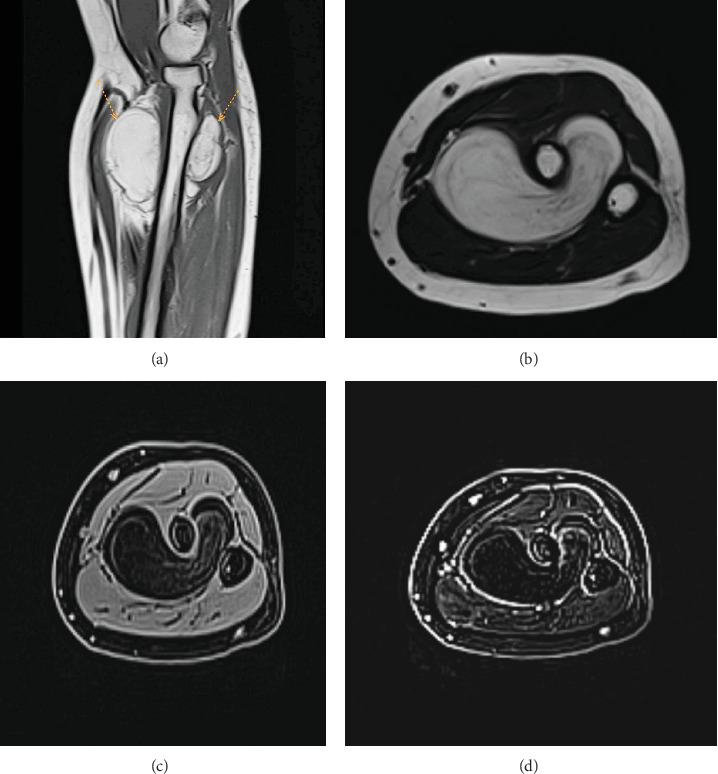 Figure 1
Peer Reviews
No public reviews on file for this paper yet. If you reviewed it on a platform where reviews are public (OpenReview, ICLR, NeurIPS, ICML), you can paste yours below so the community can read it here.
Videos
No videos yet. Explain this paper in a talk, walkthrough, or lecture? Add one.
Taxonomy
TopicsSarcoma Diagnosis and Treatment · Tumors and Oncological Cases · Musculoskeletal synovial abnormalities and treatments
1. Introduction
Lipoma is mature adipose tissue's predominant benign soft-tissue tumor [1, 2]. However, osseous lipomas are exceedingly infrequent and are categorized as intraosseous (emerging within bone) or juxtacortical (originating on bone surface). Juxtacortical lipomas can be further classified as parosteal lipomas (PLs) or sub-PLs based on their anatomical connection to the periosteum [3]. It is one of the rarest skeletal neoplasias, comprising less than 0.1% of primary bone tumors and 0.3% of all lipomas [4]. The pathogenesis of ossifying lipoma is under debate [5]. The most prevalent infected sites are the femur, humerus, proximal radius, tibia, clavicle, and pelvis. It predominantly affects adults over 40, regardless of gender [3]. Although there have been no reports of malignant changes, prompt treatment for PL seems necessary before irreversible muscle atrophy occurring, particularly in cases of nerve entrapment. The tumor is typically encapsulated and tightly adhered to the underlying periosteum, especially at sites of osseous growth. This frequently necessitates subperiosteal dissection or segmental bone excision [6].
The current report is aimed at presenting a case of PL in a 53-year-old female patient. The report is organized in line with CaReL criteria and briefly reviews the literature [7].
2. Case Presentation
2.1. Patient Information
A 53-year-old diabetic lady presented to Smart Health Tower, complaining of a slowly increasing, painless right proximal forearm mass for 3 years. For the last 2 weeks, she has felt easy fatigability and decreased grip strength after a short time of work. She reported no history of trauma.
2.2. Clinical Findings
On physical examination, a firm, immobile, painless mass was felt at the anterior proximal forearm, starting just distal to the biceps brachii tendon insertion. The range of motion of the elbow joint and the wrist joint was within normal limits. The distal pulse was positive, and there was no neurological deficit. There was no skin discoloration, scars, or dilated veins.
2.3. Diagnostic Approach
All blood tests, including complete blood count, infection markers, hemoglobin A1C (HbA1C) test, renal function, and viral markers, were within the standard limit. A computed tomography (CT) scan confirmed a well-defined fat-density mass with no bone involvement. A magnetic resonance imaging (MRI) scan showed a large, well-defined 6 × 5.5 × 3.6 cm lobulated fat-intensity space–occupying lesion. The lesion encased more than two-thirds of the proximal radial shaft, extending from the level of the radial neck and tuberosity downward. It surrounds the bone's anterior, medial, and posterior sides, traversing through the intraosseous membrane between the radius and ulna bone space. Additionally, it displaced surrounding vessels without causing intraosseous changes or invading surrounding soft tissues (Figure 1). After giving contrast, no enhancement was seen.
2.4. Therapeutic Intervention
The patient underwent surgical excision of the mass through the proximal forearm. By performing Henry's approach, the dissection was made distally between the brachioradialis and flexor carpi radialis muscles. It continued proximally between the brachioradialis muscle and the pronator teres muscle. The superficial radial nerve retracted laterally, whereas the radial artery and its accompanying venae comitantes were medially retracted after separating them from the mass. Later, the supinator muscle was separated from its broad attachment, and the mass was resected in one piece through the incising intraosseous membrane. The mass was sent for histopathological analysis, confirming the diagnosis of PL.
2.5. Follow-Up and Outcome
The postoperative period was uneventful, and the neurovascular examination was unremarkable.
3. Discussion
The present study performed a minireview of the literature to identify relevant studies on giant PLs. The literature review involved a search to identify relevant studies and was assessed based on specific criteria. A total of six reports on giant PLs were identified (Table 1). The reports spanned different locations and years, with patient ages ranging from adolescence to 83 years old. Both genders were equally affected in the reviewed cases. The presenting complaints included slowly progressive painless swellings, with one case reporting acute and progressive weakness of the right-hand extensors. Clinical examinations revealed well-circumscribed, nontender masses, often with specific characteristics such as being rubbery or immobile. Imaging analyses, including plain radiographs, ultrasounds, CT scans, and MRIs, were frequently used for diagnosis. These imaging techniques revealed various features of the masses, such as soft-tissue shadows, signal intensities indicative of lipomas, and bone-related findings. Surgical excision under general anesthesia was the management of choice in all cases. The histopathological examination (HPE) of the excised masses confirmed the diagnosis of lipoma. Postoperative outcomes were generally favorable, with no major complications or recurrences reported during follow-up.
PLs account for 0.3% of lipomas and 0.1% of primary bone tumors. They are benign soft tissue tumors, but their infiltration into skeletal muscle can resemble malignancies such as liposarcoma [1, 9]. While lipomas predominantly comprise mature adipocytes, rare instances exist where other mesenchymal elements may be present, including smooth muscle, fibrous tissue, cartilage, or bone [5]. The diaphyseal and metaphyseal regions of the long bones are the most commonly affected, and because of their deep placement, this form of tumor tends to be lazy [8]. They were first described in 1836, as reported by Fleming et al. [10]. Patients with PL have ages ranging from 3 months to the eighth decade. Between the ages of 40 and 70, patients made up roughly 50% of the cases [5]. The current case was a 53-year-old diabetic lady patient.
According to the genuine literature, the pathogenesis is still unknown, but three main theories have been outlined [5, 11]. One theory is that these tumors grow as a result of ischemia, metabolic abnormalities, or recurrent trauma, which causes pre-existing fibrous materials inside the lipoma to metaplasia and evolve into osteoblasts. Transforming growth factor (TGF)-β may have a role in ossification because it stimulates monocytes to release chemotactic and mitogenic cytokines, which attract mesenchymal and endothelial cells and encourage the creation of collagen and related components of the bone matrix [5].
These lesions typically remain asymptomatic unless complications arise from compression on a nearby neurovascular structure, resulting in neuropraxia, vessel engorgement, or limited mobility of an adjacent joint [2, 9]. The PL manifests as a firm, nontender, gradually enlarging mass over bones without fixing the skin [4]. Murugharaj et al. described a progressively enlarging right forearm mass that was presented for evaluation. The mass caused no neurological deficits but limited elbow flexion due to size and location [12]. Other scholars presented a female case with a history of a painless, progressively enlarging (approximately 4 × 3 × 2 cm) soft mass on the anterolateral aspect of her right proximal forearm. The mass limited terminal pronation [2]. In the current study, the patient presented with a three-year history of a gradually enlarging, painless mass in the right proximal forearm. The examination revealed a firm, immobile, and painless mass in the anterior proximal forearm, just distal to the biceps brachii tendon insertion.
The tumor exhibits characteristic features in imaging modalities. On CT and MRI, it appears as a homogeneous, lobulated mass adherent to the adjacent bone surface. Generally, MRI offers superior evaluation compared to CT. The tumor manifests as a juxtacortical mass with fatty signal intensity on all MRI sequences. Heterogeneity within the lesion is common, reflecting its diverse components. Cartilaginous elements are present in areas with intermediate T1 and high T2 signal intensities. Fibrovascular septa contribute to the lobulated appearance, visualized as low T1 signal intensity strands that become brighter on fat-suppressed images. Moreover, MRI excels at depicting larger areas of bone production within the lipoma. It effectively identifies adjacent muscle atrophy, a consequence of nerve entrapment, by revealing increased fatty striations within the affected muscle, particularly on T2-weighted images. In addition, MRI offers the most accurate depiction of the tumor's relationship with adjacent bone and muscle, which is crucial for surgical planning due to the firm adherence of PL to the underlying cortex at sites of bone production [8]. In the current study, MRI showed a big mass (6 × 5.5 × 3.6 cm) around most of the upper part of the radial bone. It extends from the neck to the lower part of the bone, affecting its front, middle, and back. It breached the intraosseous membrane and displaced surrounding vessels, but no intraosseous involvement or soft tissue invasion was identified. Contrast administration showed no enhancement. A CT scan confirmed it as a clear mass with no bone damage.
Surgical resection represents the definitive treatment for PL. This approach is particularly crucial in cases of nerve entrapment to prevent irreversible muscle atrophy and preserve function. During surgery, meticulous nerve dissection is paramount to avoid iatrogenic nerve injury. Unlike their soft tissue counterparts, PLs often present a surgical challenge due to their firm adherence to the underlying bone. This necessitates a more complex approach compared to the straightforward removal of juxtaposed soft tissue lipomas [4]. To separate the mass from the underlying bone, either subperiosteal dissection or segmental resection may be necessary [8]. Histological examinations often reveal disordered bone and cartilage tissue. Necrosis may or may not be visible. Integral to the diagnosis is identifying mature fatty tissue as the predominant constituent forming the tumor [5]. The mass was successfully excised via Henry's approach with no complications in the present case, and the subsequent HPE confirmed benign PL.
4. Conclusion
The tumor is a rare osseous neoplasm that may remain asymptomatic for years until it reaches a size capable of exerting pressure and causing motion difficulty. Meticulous care is paramount during surgical management to prevent iatrogenic nerve injury.
The reference list from the paper itself. Each links out to its DOI / PubMed record.
- 1Alghamdi K. Alshayie M. Large Intramuscular and Parosteal Lipoma of the Upper Limb: A Case Report and Literature Review Journal of Musculoskeletal Surgery and Research 202381758010.25259/JMSR_183_2023 · doi ↗
- 2Abdullah A. S. Ahmed A. G. Mohammed S. N. Qadir A. A. Bapir N. M. Fatah G. M. Benign Tumor Publication in One Year (2022): A Cross-Sectional Study Barw Medical Journal 202414202510.58742/wefvkv 74 · doi ↗
- 3Salama H. Kumar P. Bastawrous S. Posterior Interosseous Nerve Palsy Caused by Parosteal Lipoma: A Case Report Case Reports in Medicine 20102010378520210.1155/2010/7852022-s 2.0-8494028697820811574 PMC 2929686 · doi ↗ · pubmed ↗
- 4Chaudhary R. J. Dube V. Bhansali C. Gupta A. Balwantkar S. Parosteal Lipoma of Humerus—A Rare Case International Journal of Surgery Case Reports 20134121159116210.1016/j.ijscr.2013.09.0072-s 2.0-8489050855724252389 PMC 3860041 · doi ↗ · pubmed ↗
- 5Myint Z. W. Chow R. D. Wang L. Chou P. M. Ossifying Parosteal Lipoma of the Thoracic Spine: A Case Report and Review of Literature Journal of Community Hospital Internal Medicine Perspectives 2015512601310.3402/jchimp.v 5.260132-s 2.0-8499758941725656666 PMC 4318815 · doi ↗ · pubmed ↗
- 6Xu L. Tsang K. H. Parosteal Lipoma of the Scapula: A Case Report Hong Kong Medical Journal 2020261707210.12809/hkmj 18775932077863 · doi ↗ · pubmed ↗
- 7Prasad S. Nassar M. Azzam A. Y. Ca Re L Guideline: A Consensus-Based Guideline on Case Reports and Literature Review (Ca Re L) Barw Medical Journal 202423131910.58742/bmj.v 2i 2.89 · doi ↗
- 8Başarir K. Şahin E. Kalem M. Karaca M. O. Yildiz Y. Saglik Y. Parosteal Lipoma as a Rare Cause of Peripheral Neuropathy and Local Irritation: A Report of 12 Cases Acta Orthopaedica et Traumatologica Turcica 201751647447710.1016/j.aott.2017.02.0162-s 2.0-8503995495829128312 PMC 6197454 · doi ↗ · pubmed ↗