CT features and histogram analysis of non-contrast images for differentiating malignant and benign mediastinal lymph nodes in Non-Small Cell Lung Cancer (NSCLC)
Pakorn Prakaikietikul, Yutthaphan Wannasopha, Juntima Euathrongchit, Apichat Tantraworasin, Lorenzo Faggioni, Lorenzo Faggioni, Lorenzo Faggioni, Lorenzo Faggioni

TL;DR
This study shows that CT imaging features and histogram analysis can help distinguish between cancerous and non-cancerous lymph nodes in lung cancer patients.
Contribution
The study combines morphologic CT features and histogram analysis to improve the accuracy of diagnosing mediastinal lymph node malignancy in NSCLC.
Findings
Malignant lymph nodes were significantly larger, had irregular shapes, and showed necrotic areas.
Histogram parameters like mean attenuation, skewness, kurtosis, and entropy differed significantly between benign and malignant nodes.
Combining CT features and histogram analysis achieved an AUC of 0.870 for malignancy detection.
Abstract
To evaluate the diagnostic value of CT features and histogram analysis in distinguishing between malignant and benign mediastinal lymph nodes in patients with non-small cell lung cancer (NSCLC). This retrospective study analyzed non-contrast chest CT images from 40 NSCLC patients, comprising 80 pathology-proven mediastinal lymph nodes (46 benign, 34 metastasis). Morphologic features, including size, shape, margins, and internal composition, were independently assessed by two radiologists. Histogram analysis was conducted using the Synapse Vincent system with six parameters: mean attenuation, mean positive pixel (MPP), standard deviation (SD), skewness, kurtosis, and entropy. Statistical analysis included the Mann-Whitney test for continuous data, Fisher’s exact test for categorical data, and receiver-operating characteristic (ROC) curve analysis to assess diagnostic accuracy, with…
Genes, proteins, chemicals, diseases, species, mutations and cell lines named across the full text — each resolved to its canonical identifier and authoritative record.
Click any figure to enlarge with its caption.
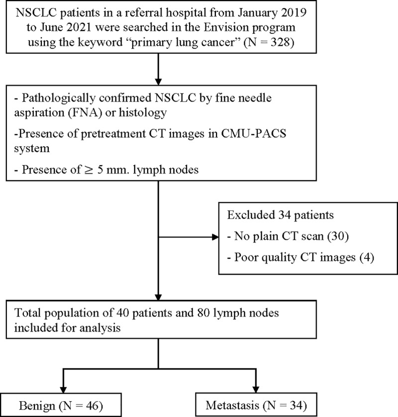 Figure 1
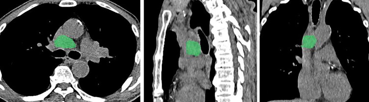 Figure 2
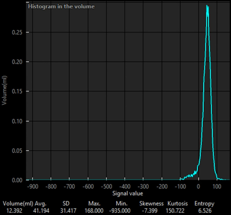 Figure 3
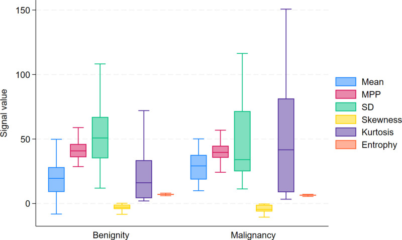 Figure 4
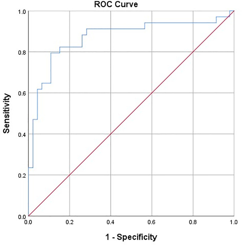 Figure 5
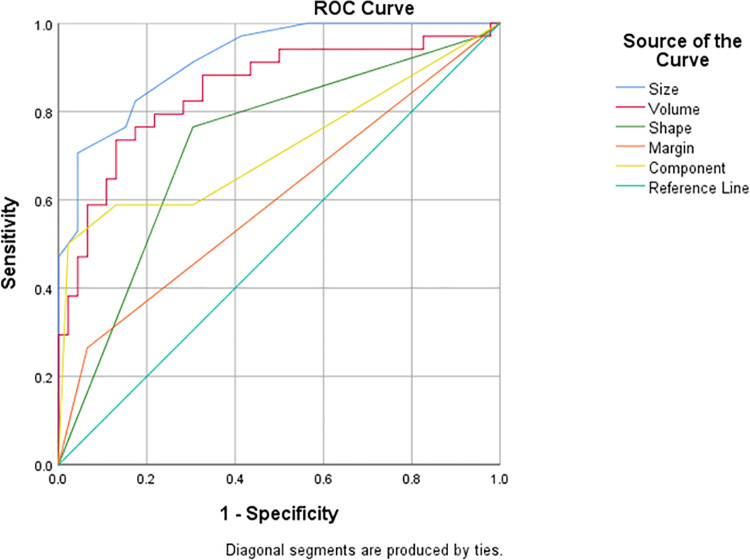 Figure 6
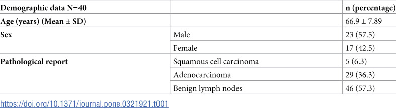 Figure 7
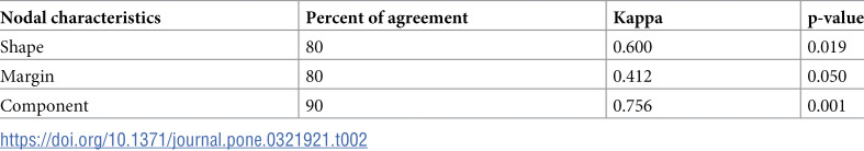 Figure 8
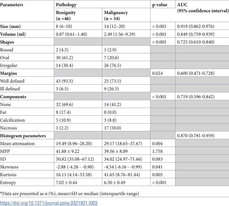 Figure 9
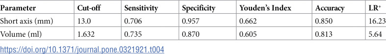 Figure 10
Peer Reviews
No public reviews on file for this paper yet. If you reviewed it on a platform where reviews are public (OpenReview, ICLR, NeurIPS, ICML), you can paste yours below so the community can read it here.
Videos
No videos yet. Explain this paper in a talk, walkthrough, or lecture? Add one.
Taxonomy
TopicsLung Cancer Diagnosis and Treatment · Radiomics and Machine Learning in Medical Imaging · Medical Imaging Techniques and Applications