Atypical Root Canal System Anatomy in a Permanent Upper First Molar: A Case Report
Slavena Georgieva, Tsvetelina Borisova-Papancheva, Denitsa Zaneva-Hristova

TL;DR
This paper presents a rare case of unusual root canal anatomy in an upper molar and details its treatment.
Contribution
The novelty lies in describing a clinical case of a C-shaped root canal configuration in a maxillary first molar.
Findings
A C-shaped root canal configuration was diagnosed in a maxillary first molar.
The case highlights the importance of adapting endodontic techniques to atypical anatomies.
Successful treatment was achieved through proper diagnosis and obturation.
Abstract
Root canal system variations can occur in each tooth group and significantly influence the outcome of the endodontic treatment. Upper first molars often present with some variations, mostly due to the presence of a second mesio-buccal root canal. Other types of atypical root canal system anatomy in upper first molars have also been reported but with a significantly smaller frequency. The aim of this article is to describe a clinical case of a C-shaped root canal configuration in a maxillary first molar - the diagnosis, preparation, irrigation, and final definitive obturation of the root canal system.
Genes, proteins, chemicals, diseases, species, mutations and cell lines named across the full text — each resolved to its canonical identifier and authoritative record.
Click any figure to enlarge with its caption.
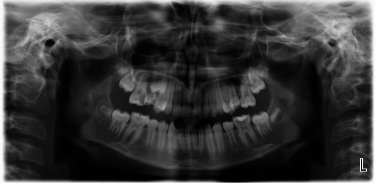 Figure 1
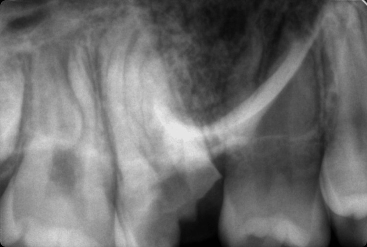 Figure 2
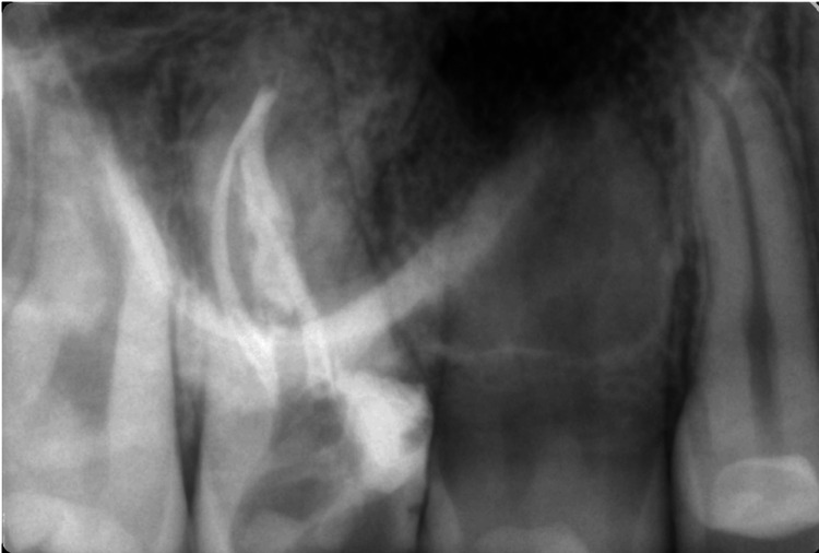 Figure 3
Peer Reviews
No public reviews on file for this paper yet. If you reviewed it on a platform where reviews are public (OpenReview, ICLR, NeurIPS, ICML), you can paste yours below so the community can read it here.
Videos
No videos yet. Explain this paper in a talk, walkthrough, or lecture? Add one.
Taxonomy
TopicsEndodontics and Root Canal Treatments · Dental Radiography and Imaging · Oral and Maxillofacial Pathology