Hippocampus and cornu ammonis: mythonyms that prevail in Terminologia Anatomica, Terminologia Neuroanatomica, and Terminologia Histologica
Jhonatan Duque-Colorado, Laura García-Orozco, Alicia Castillo-Martínez, Mariano del Sol

TL;DR
This paper examines the use and meaning of the terms 'hippocampus' and 'cornu ammonis' in anatomical terminology, highlighting inconsistencies and their mythological origins.
Contribution
The paper identifies discrepancies in terminology and clarifies the mythological etymology of hippocampus and cornu ammonis.
Findings
The terms hippocampus and cornu ammonis have mythological origins and are inconsistently used across anatomical terminologies.
Etymological analysis shows hippocampus refers to a sea horse and cornu ammonis to the horns of an Egyptian god.
The terminologies fail to meet FIPAT requirements for describing these structures.
Abstract
Julius Caesar Arantius first described the hippocampus and proposed the term hippocampum. Years later, French anatomists called the structure ram’s horns, and a decade later, it was named cornu ammonis. Although both concepts were first associated with the same structure, their use has expanded to include different but related structures. This situation can make understanding and applying the terminology more difficult. The objective of this work was to determine the presence of the terms hippocampus, cornu ammonis and their variants in Terminologia Anatomica, Terminologia Neuroanatomica, and Terminologia Histologica, evaluating their congruence in said terminologies, in addition to examining the etymology of both terms. We searched Terminologia Anatomica, Terminologia Neuroanatomica, and Terminologia Histologica for terms containing the concepts hippocampus, cornu ammonis, and their…
Genes, proteins, chemicals, diseases, species, mutations and cell lines named across the full text — each resolved to its canonical identifier and authoritative record.
Click any figure to enlarge with its caption.
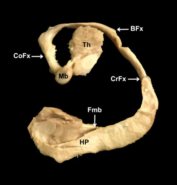 FIGURE 1
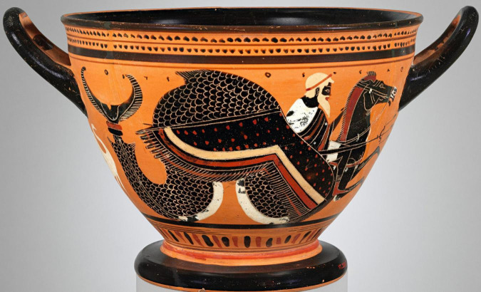 FIGURE 2
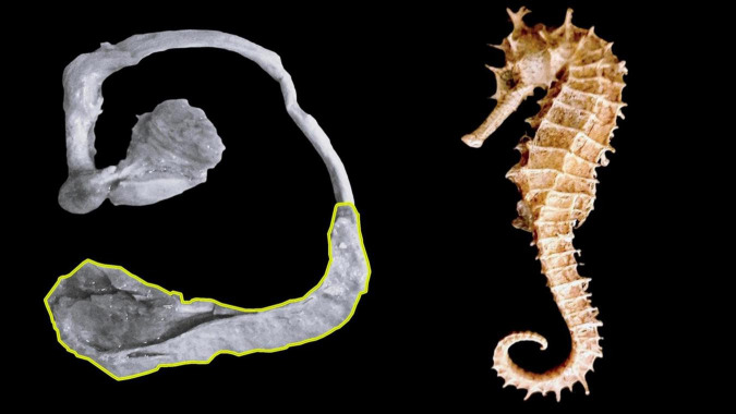 FIGURE 3
| Latin term | Latin synonym | UK English | US English | US synonym | Other | |
| 5515 | Gyrus parahippocampalis | Parahippocampal gyrus | Parahippocampal gyrus | Gyrus hippocampi | ||
| 5518 |
|
|
| Hippocampal formation | ||
| 5520 |
|
|
| |||
| 5522 | Sulcus hippocampalis | Hippocampal sulcus | Hippocampal sulcus | |||
| 5523 | Alveus hippocampi | Alveus | Alveus | |||
| 5524 | Fimbria hippocampi | Fimbria of | Fimbria of | Fimbria of fornix | ||
| 5530 | Archicortex | Archicortex | Archicortex | Archaecortex; Archecortex; Hippocampal llocortex | ||
| 5616 | Commissura hippocampi | Commissura fornicis | Hippocampal commissure | Hippocampal commissure | Commissure of fornix | Fornix transversus; Psaltarium; Lyre of David |
| 5656 | Pes hippocampi | Pes hippocampi | Pes hippocampi |
| Latin term | Latin synonym | UK English | US English | US synonym | Other | |
| 425 | Rami hippocampales | Branches to hippocampus | Branches to hippocampus | |||
| 589 | Arteriae hippocampales | Hippocampal arteries | Hippocampal arteries | |||
| 590 | Arteria hippocampalis posterior | Posterior hippocampal artery | Posterior hippocampal artery | |||
| 591 | Arteria hippocampalis media | Middle hippocampal artery | Middle hippocampal artery | |||
| 592 | Arteria hippocampalis anterior | Anterior hippocampal artery | Anterior hippocampal artery | |||
| 644 | Venae hippocampales longae | Long hippocampal veins | Long hippocampal veins | |||
| 645 | Vena hippocampalis longa anterior | Anterior long hipocampal vein | Anterior long hipocampal vein | |||
| 646 | Vena hippocampalis longa posterior | Posterior long hipocampal vein | Posterior long hipocampal vein | |||
| 2151 | Gyrus parahippocampalis | Parahippocampal gyrus | Parahippocampal gyrus | T5 | For subdivision, see Lobus limbicus. | |
| 2161 | Gyrus parahippocampalis | Parahippocampal gyrus | Parahippocampal gyrus | T5 | For subdivision, see Lobus limbicus | |
| 2164 | Verrucae hippocampi | Hippocampal warts | Hippocampal warts | |||
| 2176 |
|
|
| Inner ring of limbic lobe | ||
| 2177 | Pars precommissuralis hippocampi | Precommissural part of hippocampus | Precommissural part of hippocampus | |||
| 2178 | Pars supracommissuralis hippocampi | Supracommissural part of hippocampus | Supracommissural part of hippocampus | |||
| 2182 | Pars retrocommissuralis hippocampi | Hippocampus proprius | Retrocommissural part of hippocampus | Retrocommissural part of hippocampus | Hippocampus proper | |
| 2183 | Sulcus hippocampalis | Hippocampal sulcus | Hippocampal sulcus | |||
| 2186 | Fimbria hippocampi | Fimbria of hippocampus | Fimbria of hippocampus | |||
| 2330 |
|
|
| |||
| 2331 | Hippocampus proprius | Cornu ammonis | Hippocampus proper | Hippocampus proper | Ammon’s horn | |
| 2333 | Digitationes hippocampi | Hippocampal digitations | Hippocampal digitations | |||
| 2338 |
|
|
| |||
| 2339 | Cornu ammonis 1 | CA1 | CA1 field | CA1 field | ||
| 2340 | Cornu ammonis 2 | CA2 | CA2 field | CA2 field | ||
| 2341 | Cornu ammonis 3 | CA3 | CA3 field | CA3 field | ||
| 2342 | Cornu ammonis 3h | CA3h | CA3h field | CA3h field | CA4 | |
| 2343 |
| Strata cornus ammonis |
|
| Layers of Ammon’s horn | |
| 2351 | Interneura hippocamp | Hippocampal interneurons | Hippocampal interneurons | |||
| 2356 | Substantia alba hippocampi | Fiber connections of hippocampus | Fiber connections of hippocampus | |||
| 2358 | Neurofibrae muscosa hippocampi proprii | Mossy fibers | Mossy fibers | |||
| 2359 | Collaterales axonales hippocampi proprii | Axon collaterals of hippocampus proper | Axon collaterals of hippocampus proper | Schaffer collaterals | ||
| 2360 | Collaterales axonales hilares hippocampi | Via endofolialis | Hilar Schaffer collaterals | Hilar Schaffer collaterals | Endfolial pathway | |
| 2361 | Alveus hippocampi | Alveus | Alveus | |||
| 2362 | Fimbria hippocampi | Fimbria | Fimbria | |||
| 2365 | Commissura hippocampi | Psalterium | Hippocampal commissure | Hippocampal commissure | Psalterium | Commissura fornicis |
| 2480 | Commissura hippocampi | Psalterium | Hippocampal commissure | Hippocampal commissure | Psalterium | Commissura fornicis |
| 2587 | Area transitionis amygdalohippocampalis | Amygdalohippocampal transition area | Amygdalohippocampal transition area | |||
| 2529 | Cortex periamygdaloideus | Area transitionis amygdaloparahippocampalis | Periamygdaloid cortex | Periamygdaloid cortex | Parahippocampal amygdaloid transition area |
| Dictionary | Term | Meaning |
| A Latin dictionary (1945) | Hippocampus | -os, i, m., = ıππóκαμπoς, a sea horse: syngnathus hippocampus. |
| Cornu | Hard growth on the head of many mammals. The tooth or tusk of an elephant. Horns with the growths that represented the gods. | |
| Ammon | -onis, [Egypt, Amun] name of Jupiter, worshipped in Africa in the form of a ram (in the present-day oasis of Siwha). | |
| Oxford Latin dictionary (2012) | Hippocampus | -os, [Gk ıππóκαμπoς] a sea horse. A legendary creature associated with the gods. |
| Cornu | ||
| Ammon | -oni, m., also Hammón, Hammon, Hammon. Egyptian god with the form of a ram in Libya; he was identified as Jupiter Hammon. | |
| Perseus Digital Library (2012) | Hippocampus | -os, i, m. = ıππó-καμπoς. |
| ıππó-καμπoς | Monster with the body of a horse and the tail of a fish, on which the gods of the sea rode. | |
| Cornu | Hard, crooked growth on the head. | |
| Ammon | -onis. Egypt = ’‘Aμμων. | |
| ’‘Aμμων | 1. Originally a Libyan or Ethiopian deity; 2. Zeus, adjective of Ammonis; 3. The Greeks called him Zeus Ammon and the Romans Jupiter Ammon. |
| 1. | That, with a very limited number of exceptions, each structure shall be designated by one term only. |
| 2. | That each term in the official list shall be in Latin, each country shall being at liberty to translate the oficial Latin term into its own vernacular for teaching purposes. |
| 3. | That each term shall be, so far as possible, short and simple. |
| 4. | That the terms shall be primarily memory signs, but shall preferably have some informative or descriptive value. |
| 5. | That structures closely related topographically shall, as far as possible, have similar names. |
| 6. | That differentiating adjectives shall be, in general, arranged as opposites. |
| 7. | That eponyms shall not be used in the Official Nomenclature of Gross or Microscopic anatomy. |
| 8. | That each name must be unique. |
| 9. | That each name shall consist only of nouns and adjectives. |
| 10. | That each name shall have only one noun in nominative case. |
| 11. | That the standard word order shall have nouns following the noun they modify, and adjectives immediately following the noun they modify. |
| 12. | That nouns in genitive case are generally preferable to adjectives when the modifier means “of” an entity rather than “pertaining to” an entity.W3510 |
Peer Reviews
No public reviews on file for this paper yet. If you reviewed it on a platform where reviews are public (OpenReview, ICLR, NeurIPS, ICML), you can paste yours below so the community can read it here.
Videos
No videos yet. Explain this paper in a talk, walkthrough, or lecture? Add one.
Taxonomy
TopicsMedical and Biological Sciences
Introduction
The hippocampus, located deep in the temporal lobe and surrounded by the cerebral ventricles, plays a fundamental role in emotional regulation, learning, memory, and various cognitive functions (White et al., 2024). The Italian anatomist Julius Caesar Arantius (1530-1589) first described it in the first chapter of his work De humano foetu liber (Arantius, 1587), where he proposed the term hippocampum due to its similarity to a sea horse. Years later, French anatomists visualized that the hippocampus, macroscopically, presented a different shape to that of a sea horse, which is why it was initially called ram’s horns (Winslow, 1732), and a decade later, it was named cornu ammonis (Garengeot, 1742). Since then, the terms hippocampus and cornu ammonis have become central to medical literature. Initially, both concepts referred to the same structure, but their usage has expanded to include distinct yet related formations (Anand and Dhikav, 2012; Twait et al., 2023; White et al., 2024), a circumstance that can complicate the understanding and function of the structure.
To avoid confusion about the structures that make up the human being, the International Federation of Associations of Anatomists (IFAA) has implemented several programs, including the Federative International Programme for Anatomical Terminology (FIPAT). The latter provides a set of terminologies that are accepted worldwide for the description of anatomical structures. Morphological Sciences use these terminologies as linguistic tools that allow the reader to interpret the content accurately and unambiguously (Duque-Colorado et al., 2023). Thus, the objective of this work was to determine the presence of the terms hippocampus, cornu ammonis and their variants in Terminologia Anatomica (FIPAT, 2019), Terminologia Neuroanatomica (FIPAT, 2017) and Terminologia Histologica (Federative International Committee on Anatomical Terminology [FICAT], 2008), evaluating their congruence in said terminologies, in addition to analyzing the etymology of both terms and their concordance with the guidelines established by FIPAT.
Materials and methods
Review and etymological analysis
We conducted a search in Terminologia Anatomica (FIPAT, 2019), Terminologia Neuroanatomica (FIPAT, 2017), and Terminologia Histologica (Federative International Committee on Anatomical Terminology [FICAT], 2008) for terms containing the concepts hippocampus, cornu ammonis, and their derivatives. The terms hippocampus and cornu ammonis underwent review and etymological analysis using a Latin dictionary (Lewis and Short, 1945), the Oxford Latin Dictionary (Oxford Dictionaries, 2012), and the Perseus Digital Library (Crane, 1987). The analysis included a review of the Regular Anatomical Terminology (RAT) rules to assess the consistency of the terms with the guidelines and objectives set by FIPAT.
Dissection
One cerebral with no macroscopic evidence of disease, belonging to a male adult, was fixed in 10% formalin, after 8 weeks the hemisphere was washed for several hours in fresh water. To preserve the white matter (fornix, anterior column of the fornix and mammillothalamic tract) we used the Klingler method (Castruita et al., 2024). The arachnoid, pia mater and vascular structures were removed, and the hemisphere was placed in a plastic bag and stored in a deep freezer at –20°C for 4 weeks. The hemisphere was thawed at room temperature and washed in running water. Once thawed and during dissections, the samples were preserved in a 70% ethanol solution.
The cortex of the subrostral area, including the paraterminal and paraolfactory gyrus, was carefully removed. Removal of the cingulate gyrus cortex exposed the supracommissural part of the hippocampus and its lateral longitudinal striae on the upper surface of the corpus callosum. The corpus callosum was dissected from the genu to the splenium, along with the septum pellucidum, allowing visualization of the upper surface of the thalamus and the body of fornix.
A No. 10 blade then made cuts along the superior margin of the caudate nucleus superiorly, the isthmus of cingulate gyrus posteriorly, the cerebral peduncle inferiorly, and the posteromedial orbital gyri anteriorly. Then, in a lateral to medial direction, the cortex was removed along with the superficial U-shaped fibers of the insula, identifying the thalamus on the lateral side and the fornix and parahippocampal gyrus on the medial side. Using a No. 15 blade, the white matter of the temporal peduncle was cut approximately 5 mm below the inferior limiting sulcus, identifying the alveus and collateral eminence. The fornix was dissected gradually, starting at the level of the anterior commissure down to the fimbria.
Using a No. 10 blade, we cut the anterior part of the temporal peduncle at the level of the amygdaloid body, allowing identification of the parahippocampal/hippocampal gyrus complex from different angles. In the final step, we dissected the hippocampus from the parahippocampal gyrus along the hippocampal sulcus.
Results
In Terminologia Anatomica (FIPAT, 2019), the term hippocampus was detailed with code 5518 as a general concept, with its synonym in US English corresponding to “hippocampal formation,” which encompassed several structures. Similarly, several related concepts were present, including the term hippocampus proper, a Latin synonym for the primary term cornu ammonis with code 5520, the elements of which are presented in Table 1.
Terminologia Neuroanatomica (FIPAT, 2017) did not include the term hippocampus. However, this terminology included more concepts derived from the term in question. Repeated concepts identified with different codes included formatio hippocampi, appearing under codes 2176 and 2330. Another term was hippocampus proprius, which was detailed as a Latin synonym represented by code 2182, while code 2331 represented a primary term in Latin, whose synonym was cornu ammonis. Regarding cornu ammonis, in addition to being a Latin synonym for hippocampus proprius, it corresponded to several primary terms in Latin cornu ammonis 1, cornu ammonis 2, cornu ammonis 3, and cornu ammonis 3h, identified with codes 2339, 2340, 2341, and 2342, respectively. Their Latin synonym, as well as their UK and US English equivalents, used the acronym CA. The details of this paragraph appear in Table 2.
In Terminologia Histologica (Federative International Committee on Anatomical Terminology [FICAT], 2008), under code H3.11.03.6.01114, the term hippocampus proprius or cornu ammonis was identified as the primary term in Latin.
According to different texts, as shown in Table 3, the term hippocampus from the Greek íππóκαμπoς, refers to a sea horse, which is also a mythological creature. On the other hand, the term cornu ammonis was reviewed separately, with cornu referring to the protuberances on the head that depicted the gods. Along the same lines, ammonis was identified as Ammon, derived from the Greek ’‘Aμμων, the name of a god venerated in Africa, whose cult spread to Greece.
The term hippocampus etymologically refers to a sea horse. However, during the dissection performed (Figure 1), we observed that the hippocampus does not have this form. For the hippocampus to resemble a sea horse, it must incorporate the fornix. Thus, this term does not comply with numeral four of the RAT rules, which, in turn, causes the words that include or derive from the term hippocampus to fail to meet the RAT rules, as outlined in Table 4. Along these lines, the term cornu ammonis refers to the horns of an African god called Ammon, making it an eponym with a mythological background. Such a term violates number seven and goes against the purposes of FIPAT.
Dissection of hippocampus and fornix. HP, hippocampus. Fmb, fimbria. CrFx, crus of fornix. BFx, body of fornix. CoFx, column of fornix. Mb, mammillary body. Th, thalamus.
Discussion
“Horum ventriculorum basi, quae intrb ad medium respicit, candida insurgens supereminet, and quasi adnascitur substantia, quz ab inferiori superficie, uelut additamentum extollitur, psalloidique corpori, seu testudini est continua, ac per longitudinem, in anteriora, uersus frontem protenditur inaequalique, ac flexuosa figura praedita est, quae Hippocampi, hoc est marini equuli effigiem rcfert, vel potius, bombycini uernis candidi spinalis medulle initium hinc inde amplexantis” (Arantius, 1587).
The above sentence corresponds to the first description of the hippocampus by Julius Caesar Arantius, considered by Lewis (1923) to be the worst anatomical description in existence. This is because it does not clarify whether the comparison refers to a fish or a beast, and it prevents us from determining with certainty which of its ends represents the head.
Arantius (1587) observation was largely forgotten until two centuries after the publication of the Eustachian tables (Eustachi, 1714) when it regained attention and led to the introduction of the terms ram’s horns (Winslow, 1732) and cornu ammonis horns (Garengeot, 1742). This last term replaced the term ram’s horns without any sound reason, and although it was adopted by several anatomists of the time, Hyrtl (1880) harshly criticized this term and ridiculed it, stating the following: “In order that the organ of the human soul should have no ordinary horns, out of the Cornua arietis were made the Cornua Ammonis, which amount to the same thing.” Later, the hippocampus received more names in different languages, such as gerollte wulst “rolled lump” and kolbe “mass-like structure” (Meyer, 1971). It was even confused with hippopotamus by Mayer (1779), and this term was adopted by several German anatomists. Thus, the term hippocampus exemplifies how creativity influences the assignment of names, prioritizing ingenious concepts over precise scientific descriptions (Burdach, 1822).
The term hippocampus, with code 5518, is only present in Terminologia Anatomica (FIPAT, 2019). Its absence in Terminologia Histologica (Federative International Committee on Anatomical Terminology [FICAT], 2008) was to be expected since it is a macroscopic structure. Its absence in Terminologia Neuroanatomica (FIPAT, 2017) may be because the US synonym for hippocampus in Terminologia Anatomica (FIPAT, 2019) is hippocampal formation, a concept listed as the primary term in Terminologia Neuroanatomica (FIPAT, 2017) under codes 2176 and 2330. Therefore, hippocampus may be equivalent to formatio hippocampi (hippocampal formation). However, several studies indicate that the hippocampus is a different structure from the hippocampal formation (Jarrard and Davidson, 1991; Anand and Dhikav, 2012; Schultz and Engelhardt, 2014; Pang et al., 2019).
The hippocampus is a structure comprising the hippocampus proper and the dentate gyrus, both regions coiled around each other (Tatu and Vuillier, 2014). The hippocampus proper is structured mainly from pyramidal neurons, and the dentate gyrus from granule neurons. The hippocampus proper is structured mainly from pyramidal neurons, and the dentate gyrus from granule cells. This region is a site of neurogenesis, particularly in the dentate gyrus, so this structure contributes mainly to its role in learning, memory (Fabiano et al., 2025), time perception (Tatu and Vuillier, 2014), and emotional memory (Maggio and Segal, 2012). On the other hand, the hippocampal formation includes several interconnected regions, namely the hippocampus (hippocampus proper and dentate gyrus), subiculum, and entorhinal cortex (Pang et al., 2019). In this sense, the hippocampal formation covers a broader range of functions related to memory and spatial navigation since the subiculum allows processing and amplifying hippocampal information while the entorhinal cortex integrates and channels cortical information, thus creating a bidirectional flow for memory consolidation and recovery (Böhm et al., 2017; Roy et al., 2017). Consequently, the entorhinal cortex is a key information input and output center (Fabiano et al., 2025).
Considering the above mentioned, there are deficiencies in the terminologies, thinking that if hippocampus and hippocampal formation refer to the same thing, there would be no agreement between Terminologia Anatomica (FIPAT, 2019) and Terminologia Neuroanatomica (FIPAT, 2017), since the same structure has two different names, an aspect that goes against the first numeral of the RAT rules, so a single concept should be considered as the primary term in Latin, being hippocampus or formatio hippocampi. If these are distinct structures, the US synonym must be discarded, as they are referred to by the same name, which contradicts section eight of the RAT rules.
In this same context, another element that generates discrepancies between terminologies is that in Terminologia Anatomica (FIPAT, 2019), under code 5520, the primary term is found in Latin cornu ammonis, with a synonym in Latin hippocampus proper. A completely different condition appears in Terminologia Neuroanatomica (FIPAT, 2017), where under code 2331, it is designated as the primary term in Latin, hippocampus proprius, with the Latin synonym cornu ammonis. This aspect explains the lack of synergy between terminologies, as the same structure is referred to by different names in documents that belong to the same program, such as FIPAT, which aims to serve as a reference for the identification and standardized naming of human structures in the medical and morphological fields. Likewise, for each terminology, the term used in UK and US English is derived from the primary Latin term, resulting in increased confusion in the morphological and educational fields, as scientific literature is predominantly written and published in English.
In Terminologia Neuroanatomica (FIPAT, 2017), the regions of the hippocampus proper are given by several subfields, which are detailed as main terms in Latin by cornu ammonis 1, cornu ammonis 2, cornu ammonis 3 and cornu ammonis 3h. Despite this, their synonyms in Latin, translated into UK and US English, are CA1, CA2, CA3, and CA3h, respectively, acronyms that do not provide references about the structure they represent lack informative value (Duque-Colorado et al., 2024), which does not promote the purposes of FIPAT and goes against the RAT rules.
Arantius (1587) proposed the term hippocampum for this brain structure because of its similarity to a sea horse. Our results indicated that the term hippocampus, which comes from the Greek íππóκαμπoς, arises from the union of íππoς (horse) and καμπoς (sea monster), so although it is associated with a sea horse, it has also been associated with a mythical monster with a fishtail and a horse’s body, on which the sea god Poseidon rode (Figure 2). Thus, the term hippocampus corresponds to a zoonym with a mythological background. Despite the above, we agree with some authors (Lewis, 1923; Winslow, 1732; Garengeot, 1742) that the hippocampus at a macroscopic level does not share the same shape as a seahorse, since for them to compare, in addition to considering the hippocampus, the structures that make up the fornix should also be considered (Figure 3).
Attic vase painted by the Leagros group in the 6th century BCE, depicting Poseidon riding a hippocampus.
Comparison between hippocampus and seahorse. (A) Hippocampus highlighted in yellow. (B) Seahorse.
Although initially, Garengeot (1742) used the term cornu ammonis to refer to the hippocampus, it is currently considered a structure that is part of the hippocampus, according to Terminologia Anatomica (FIPAT, 2019) or of the formation hippocampi according to Terminologia Neuroanatomica (FIPAT, 2017). According to the description made by Garengeot (1742), the hippocampus had the shape of a ram’s horns, which is why he called it cornu ammonis (Ammons’ horns). The etymology of the words tells us that cornu refers to protuberances that protrude from the head and are associated with deities (Lewis and Short, 1945). This is because, over time, art and myths have preserved animal parts, such as wings, hooves, and horns, for the gods, with artists using horns as a symbol of power. In this sense, the term cornu ammonis, in addition to representing a structure of the hippocampus, refers to the horns of the god Ammon (’‘Aμμων), a supreme deity in Libya and Egypt. Initially, this god appeared with antlers on his head. His depiction changed to ram’s horns when his cult spread to Greece in the 5th century BCE, where he became known as Zeus-Ammon. This suggests that this term is based on an eponym in a mythological context, making it a mythonym.
Currently, the term cornu ammonis refers to the regions of the hippocampus, which are visible microscopically but not macroscopically. These fields are classified based on the characteristics of the neurons that compose each region; therefore, Terminologia Anatomica (FIPAT, 2019) should not list them.
Hippocampus and cornu ammonis are not the only terms used that barely make sense. In Terminologia Anatomica (FIPAT, 2019), criticisms have been made of different terms, such as diastema (Panes and del Sol, 2020), humor aquosus (García-Orozco et al., 2023), fossa and fovea (Alarcón-Apablaza et al., 2024), calva (García-Orozco et al., 2024), among other terms. For its part, we consider that Terminologia Neuroanatomica (FIPAT, 2017) has received less attention. However, terms included in this document, such as splenium (Pearce, 2008), the structures that make up the brainstem (Watson et al., 2019), fornix (Dogan et al., 2022), thalamus (García-Cabezas et al., 2021), corpus amygdaloideum (Maldonado-Rengel et al., 2021), substantia chromatophilca (Duque-Colorado et al., 2023) and neuron parvum fluorescens (Valenzuela-Aedo et al., 2024) have also been subject to criticism. Morphological Sciences, Medical Sciences, and Neuroscience are constantly evolving; therefore, it is natural that FIPAT terminologies undergo continuous transformation. Thus, we believe it is appropriate to gradually replace terms unrelated to the structures to which they refer and that the terms included be governed by the 12 principles established in the RAT rules, thus allowing for the construction of contemporary terminologies.
The RAT rules consist of a series of recommendations whose application ensures the use of precise terms for new and known structures. This approach aligns with the objective of the FIPAT terminologies, ensuring accurate and well-defined concepts that facilitate communication among health professionals and enhance teaching-learning processes in morphological areas (Duque-Colorado et al., 2024). Therefore, although various terms have spread and consolidated in the medical, morphological, and teaching fields, we consider it pertinent to re-evaluate the terms mentioned in this chapter.
The first descriptions of human body structures by anatomists were fundamental to the development of modern medicine. Through detailed studies and precise observations, these pioneers not only described previously unknown structures but also laid the foundations for Terminologia Anatomica (FIPAT, 2019), Terminologia Neuroanatomica (FIPAT, 2017), and Terminologia Histologica (Federative International Committee on Anatomical Terminology [FICAT], 2008). While some of these terms have been subject to criticism and revision over time, driven by advances in scientific and cultural understanding, their value and original contribution remain significant. The work of these anatomists not only bequeathed a fundamental structure of knowledge but also highlighted the importance of rigorous observation and systematization of the human body—principles that remain key pillars of medical research today.
Conclusion
There is no congruence between terminologies to designate the terms hippocampus and cornu ammonis. Both terms are mythonyms, although they originate from different myths, hippocampus relates to a sea monster, while cornu ammonis refers to an Egyptian god. Although the hippocampus was initially named as such because of its similarity to a seahorse, dissection has shown that it does not have the shape of this marine animal. Consequently, both terms contradict the purposes and objectives of FIPAT. Therefore, advancing further studies in this area would be relevant to implement the necessary modifications in these documents, which regulate uniform communication among health professionals, morphologists, neuroscience professionals, and students.
The reference list from the paper itself. Each links out to its DOI / PubMed record.
- 1Alarcón-Apablaza J.García-Orozco L.Duque-Colorado J.Fuentes R. (2024). Unifcation of the term fossa and fovea in Terminologia Anatómica. Int. J. Morphol. 42 9–16. 10.4067/S 0717-95022024000100009 27315006 · doi ↗
- 2Anand K. S.Dhikav V. (2012). Hippocampus in health and disease: An overview. Ann. Indian Acad. Neurol. 15 239–246. 10.4103/0972-2327.104323 23349586 PMC 3548359 · doi ↗ · pubmed ↗
- 3Arantius J. C. (1587). De Humano foetu liber tertio editus, ac Recognitus. Anatomicarum Observationum liber, ac de Tumoribus Secundum Locos Affectos Liber. Venice: Jacob Brechtanus.
- 4Böhm C.Peng Y.Geiger J. R. P.Schmitz D. (2017). Routes to, from and within the subiculum. Cell Tissue Res. 373 557–563. 10.1007/s 00441-018-2848-4 29808383 · doi ↗ · pubmed ↗
- 5Burdach K. F. (1822). Vom Baue und Leben des Gehirns. Leipzig: Dyk’sche Buchhandlung.
- 6Castruita R.Perez S.Baldoncini M.Forlizzi V.Martínez A. (2024). The frontal aslant tract: Anatomical description and case report. Surg. Neurol. Int. 15:397. 10.25259/SNI_809_2024 39640333 PMC 11618791 · doi ↗ · pubmed ↗
- 7Crane G. R. (1987). Perseus Digital Library. Medford, MA: Tufts University.
- 8Dogan E.Gungor A.Dogulu F.Türe U. (2022). The historical evolution of the fornix and its terminology: A review. Neurosurg. Rev. 45 979–988. 10.1007/s 10143-021-01635-w 34498223 · doi ↗ · pubmed ↗