Protocol for label-free quantitative lysine lactylproteome profiling
Yu Ran, Qiqing Yang, Lianqi Zhou, Heyu Li, Bing Yang, Long Zhang

TL;DR
This paper describes a detailed protocol for analyzing lactylation, a protein modification linked to L-lactate, using label-free mass spectrometry techniques.
Contribution
The paper introduces a comprehensive, step-by-step protocol for profiling and quantifying the lactylproteome without the need for labeling.
Findings
The protocol includes cell preparation, protein extraction, and lactylpeptide enrichment using antibody-conjugated agarose beads.
LC-MS/MS parameters and MaxQuant settings are provided for label-free quantification of lactylated peptides.
The method enables global profiling of lactylation, a PTM involved in cellular signaling and metabolism.
Abstract
L-lactate has been recognized as an essential molecule for signaling and metabolic balance. Lactylation, a post-translational modification (PTM) derived from L-lactate, is commonly observed on various proteins and plays essential roles in cellular processes. Here, we present a protocol to globally profile the lactylation proteome and perform label-free quantification. We provide steps for cell preparation, protein extraction, digestion, peptide desalting, and lactylpeptide enrichment. Additionally, we outline the parameters for liquid chromatography-tandem mass spectrometry (LC-MS/MS) analysis. For complete details on the use and execution of this protocol, please refer to Li et al.1 •Instructions for the preparation of cell culture, transfection, and lactate treatment•Steps for enriching lactylpeptide based on antibody-conjugated agarose beads•Guidance on LC-MS setup and MaxQuant…
Genes, proteins, chemicals, diseases, species, mutations and cell lines named across the full text — each resolved to its canonical identifier and authoritative record.
Click any figure to enlarge with its caption.
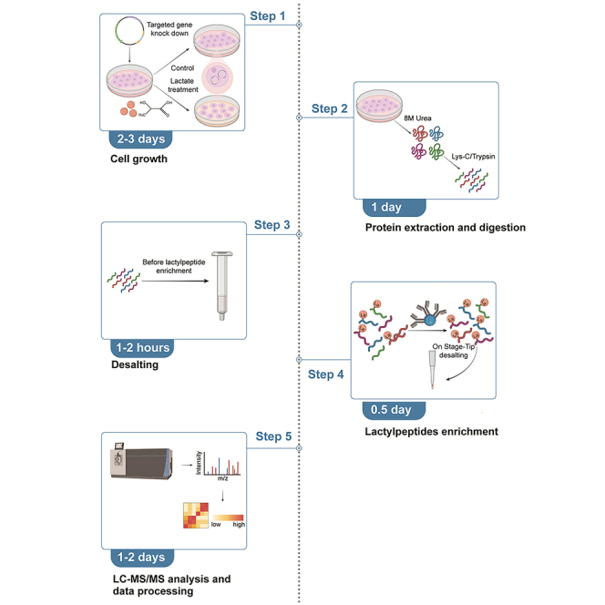 Figure 1
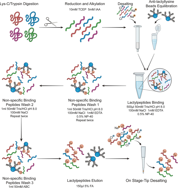 Figure 2
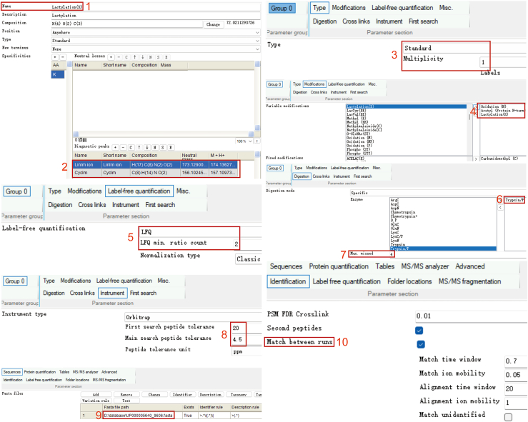 Figure 3
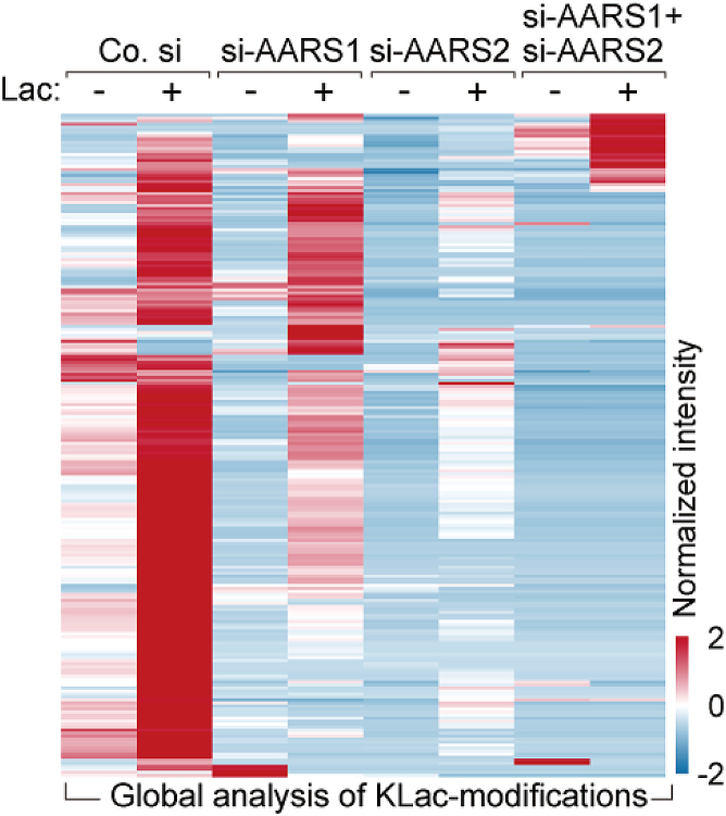 Figure 4
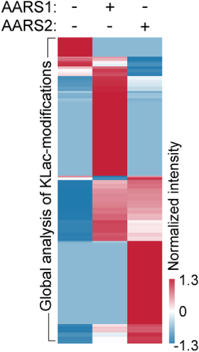 Figure 5
Peer Reviews
No public reviews on file for this paper yet. If you reviewed it on a platform where reviews are public (OpenReview, ICLR, NeurIPS, ICML), you can paste yours below so the community can read it here.
Videos
No videos yet. Explain this paper in a talk, walkthrough, or lecture? Add one.
Taxonomy
TopicsAdvanced Proteomics Techniques and Applications · Mass Spectrometry Techniques and Applications · RNA and protein synthesis mechanisms
Before you begin
Preparation
This protocol describes quantitative analysis of protein lactylation in AARS1 and AARS2 (AARS1/2) knock down, AARS1/2 overexpression and wild type cells. For experimental uses, following materials should be prepared.
- 1.Synthesize mammalian cell expression vectors encoding AARS1/2 and siRNAs targeting AARS1/2.
- 2.Culture HEK293T cells (ATCC 3216) and HeLa cells (ATCC CCL-2) in DMEM (Fisher Scientific, Cat#31966047) supplemented with 10% (v/v) of fetal bovine serum (FBS Gibco, Cat#10270106), 100 mg/mL penicillin and streptomycin (Invitrogen, Cat#15140-122).
- 3.Maintain cell lines in a humidified incubator at 37°C, with 5% CO_2_. Regularly check for mycoplasma contamination using the EZ PCR Mycoplasma detection Kit (Biological Industries, Cat#20-700-20).
- 4.Refresh the cell medium at least every 48 h, by aspirating half of the old medium (taking care not to alter the spheroids) and adding an equal volume of fresh medium.
- 5.Calibrate LC-MS equipment and run quality control samples such as HeLa cell lysate standards to check if the LC-MS/MS performs as expected.
Key resources table
REAGENT or RESOURCESOURCEIDENTIFIERChemicals, peptides, and recombinant proteinsDulbecco’s phosphate-buffered saline (PBS) (1 ×)Thermo Fisher ScientificCat# 14040091Dulbecco’s modified Eagle’s medium (DMEM)Thermo Fisher ScientificCat# 31966047Trypsin EDTA (0.05%)Thermo Fisher ScientificCat# 25300054Fetal bovine serum (FBS)GibcoCat# 10270106Penicillin-Streptomycin (10,000 U/mL)InvitrogenCat# 15140-122L-Lactic AcidAladdinCat# A2205202Dimethyl sulfoxide (DMSO)VWRCat# A3672Ammonium bicarbonateCayman ChemicalCat# 16894Trypsin Gold, mass spectrometry gradePromegaCat# V5280C18 Solid phase extraction disksEmporeCat# 2215-C1833 μm Polymeric sorbent solid-phase extraction cartridgesStrata-XCat# 8B-S100-AAKAnti-L-lactyllysine antibody-conjugated agarose beadsPTM BioCat# PTM-1404Tris (2-carboxyethyl) phosphinehydrochloride (TCEP)Sigma-AldrichCat# 24615-84-7Iodoacetamide (IAA)Sigma-AldrichCat# 144-48-9Formic acid (FA), LC-MS gradeThermo Fisher ScientificCat# 28905Lys-C, mass Spec gradePromegaCat# VA117ATrifluoroacetic acid (TFA), eluent additive for LC-MSSigma-AldrichCAS# 76-05-1Tris-HCl (tris(hydroxymethyl)aminomethane hydrochloride)VWRCAS# 1185-53-1Urea, high purityVWRCAS# 57-13-6Acetonitrile (ACN), Optima LC/MS gradeThermo Fisher ScientificCAS# A955-500Methanol, Optima LC/MS gradeThermo Fisher ScientificCAS# 67-56-1cOmplete, EDTA-free Protease Inhibitor CocktailRocheCAS# 04693132001Deacetylase Inhibitor CocktailMCECAS# HY-K0030Critical commercial assaysPolyethylenimineSigma-AldrichCAS# 9002-98-6Pierce BCA Protein Assay KitThermo Fisher ScientificCat# 23225Lipofectamine RNAiMAX Transfection1070 ReagentThermo Fisher ScientificCat# 13778150EZ PCR Mycoplasma detection KitBiological IndustriesCat# 20-700-20Experimental models: Cell linesHuman cell line, HEK293TATCCCat# CRL-3216Human cell line, HeLaATCCCat# CRM-CCL-2OligonucleotidesHuman AARS1 siRNA #1 5′–3′-sequence GCAGTGAGATCCACTACGARibo Life ScienceN/AHuman AARS1 siRNA #2 5′–3′-sequence GTTTGGCATTCCCATTGAARibo Life ScienceN/AHuman AARS2 siRNA #1 5′–3′-sequence GGAACCTGGTCTTCATGCARibo Life ScienceN/AHuman AARS2 siRNA #2 5′–3′-sequence GTTTGGCATTCCCATTGAARibo Life ScienceN/ANegative control siRNARibo Life ScienceN/ARecombinant DNApLV-AARS1-MycN/AN/ApLV-AARS2-MycN/AN/ASoftware and algorithmsMaxQuantTyanova et al.2MaxQuant v2.0.3.0Microsoft ExcelMicrosoft CorporationN/AUniProt database humanN/Ahttps://www.uniprot.org/proteomes/UP000005640OtherCO_2_ incubatorThermo Fisher ScientificCat#51033770SpeedVac vacuum concentratorsThermo Fisher ScientificCat#SPD111V-115Benchtop freeze dryersLabconcoCat# 10-400-021MicrocentrifugeBMG LABTECHCat# 22620667Nunc EasYDish dishesThermo Fisher ScientificCat# 150464Axygen 1.5 mL MaxyClear SnaplockAxygenCat# MCT-150-CAxygen 1000 μL pipet tipsAxygenCat# T-1000-BAxygen 10 μL microvolume pipet tipsAxygenCat# T-300-R-SAxygen 200 μL Universal Fit pipet tipAxygenCat# T-200-CThermo Scientific Exploris 480 mass spectrometerThermo Fisher ScientificCat# BRE725533Thermo Scientific EASY-nLC 1200 instrumentThermo Fisher ScientificCat# LC140
Materials and equipment
Cell Culture MediumReagentFinal concentrationAmountDMEMN/A500 mLFetal Bovine Serum10%50 mLPenicillin/ streptomycin (10,000 U/ mL)100 U/ mL5 mLTotalN/A555 mL Note: Cell Culture Medium can be stored at 4°C for 1 month. Denaturing Lysis BufferReagentFinal concentrationAmountUrea8 M240 mgTris/HCl pH 8.5 (1 M)100 mM50 μLLC-MS grade H2ON/A270 μLProtease Inhibitor (100×)N/A5 μLDeacetylase Inhibitor (100×)N/A5 μLTotalN/A500 μL Note: Urea is unstable. Prepare freshly before each use. Lactylpeptide Binding bufferReagentFinal concentrationAmountTris/HCl pH 8.0 (1 M)50 mM2.5 mLNaCl (5 M)100 mM1 mLEDTA (500 mM)1 mM100 μLNP-40 (20%)0.5% (v/v)1.25 mLLC-MS grade H_2_ON/A45.15 mLTotalN/A56 mL Note: Lactylpeptide Binding buffer can be stored at room temperature for 1 month. Lactylpeptide Wash bufferReagentFinal concentrationAmountTris/HCl pH 8.5 (1 M)50 mM2.5 mLNaCl (5 M)100 mM1 mLLC-MS grade H_2_ON/A46.5 mLTotalN/A50 mL Note: Lactylpeptide Wash buffer can be stored at room temperature for 1 month. Lactylpeptide Elution bufferReagentFinal concentrationAmountFA5% (v/v)500 μLLC-MS grade H_2_ON/A10 mLTotalN/A10 mL Note: As the concentration of FA is relatively high, Lactylpeptide Elution buffer can be stored at room temperature for 1 week. Ammonium Bicarbonate BufferReagentFinal concentrationAmountAmmonium Bicarbonate400 mM1.56 gLC-MS grade H_2_ON/A50 mLTotalN/A50 mL Note: 400 mM ammonium bicarbonate (ABC) buffer stock can be stored at room temperature for 1 month. Dilute ABC buffer to 50 mM before use. Desalting activation bufferReagentFinal concentrationAmountLC-MS grade Methanol100% (v/v)10 mLTotalN/A10 mL Note: Methanol is highly flammable and harmful if swallowed. Keep away from heat, hot surfaces, sparks, open flames and other ignition sources. Note: Desalting activation buffer can be stored at room temperature for 1 month. SPE column equilibration bufferReagentFinal concentrationAmount40% TFA0.1% (v/v)25 μLLC-MS grade H_2_ON/A9.975 mLTotalN/A10 mL Note: TFA is highly corrosive and volatile. Handle in a fume hood and wear gloves and goggles when operate experiments. Note: SPE column equilibration buffer can be stored at room temperature for 1 month. SPE elution bufferReagentFinal concentrationAmountACN80% (v/v)8 mL40% TFA0.1% (v/v)25 μLLC-MS grade H_2_O19.9% (v/v)1.975 mLTotalN/A560 mL Note: TFA is highly corrosive and volatile. ACN is highly flammable. Handle in a fume hood and wear gloves and goggles when operate experiments. Check pH before use and ensure pH is below 1. Note: SPE elution buffer can be stored at room temperature for 1 month. Stage tip desalting buffer AReagentFinal concentrationAmountFA0.1% (v/v)10 μLLC-MS grade H_2_ON/A9.99 mLTotalN/A10 mL Note: Stage tip desalting buffer A can be stored at room temperature for 1 month. Stage tip desalting buffer BReagentFinal concentrationAmountFA0.1% (v/v)10 μLACN99.9% (v/v)9.99 mLTotalN/A10 mL Note: Stage tip desalting buffer B can be stored at room temperature for 1 month. Stage tip desalting buffer CReagentFinal concentrationAmountACN40% (v/v)4 mLFA0.1% (v/v)10 μLLC-MS grade H_2_O59.9% (v/v)5.99 mLTotalN/A10 mL Note: Stage tip desalting buffer C can be stored at room temperature for 1 month. LC mobile phase AReagentFinal concentrationAmountACN2% (v/v)500 μLFA0.1% (v/v)25 μLLC-MS grade H_2_O97.9% (v/v)24.975 mLTotalN/A25 mL LC mobile phase BReagentFinal concentrationAmountACN80% (v/v)20 mLFA0.1% (v/v)25 μLLC-MS grade H_2_O20% (v/v)4.975 mLTotalN/A25 mL Note: Degassing of LC mobile phase for 30 min using ultrasound. Note: LC mobile phase can be stored at room temperature for 1 week. CRITICAL: The concentration of ACN should not greater than 95%, otherwise it will damage the HPLC instrument. Reduction buffer (TCEP buffer)ReagentFinal concentrationAmountTCEP1 M2.50187 gLC-MS grade H_2_ON/A10 mLTotalN/A10 mL Alkylation buffer (IAA buffer)ReagentFinal concentrationAmountIAA500 mM924.82 mgLC-MS grade H_2_ON/A10 mLTotalN/A10 mL Note: Reduction and Alkylation buffer can be stored at −20°C for 1 month. L-lactic acid stockReagentFinal concentrationAmountL-lactic acid10 M9.008 gMilliQ waterN/A10 mLTotalN/A10 mL Note: L-lactic acid stock can be stored at −20°C for 1 month.
Step-by-step method details
Cell culture
Timing: 2 days
This part describes the preparation of a HEK293T cell culture for transfection.
- 1.Pre-warm DMEM, PBS and trypsin-EDTA in a 37°C water bath.
- 2.Cultivate HEK293T cells in a 10 cm dish under humidified conditions with 5% CO_2_ at 37°C using DMEM medium until they reach 80%–90% confluency.
- 3.Aspirate the growth medium and gently wash the cells twice with 2 mL of sterile PBS.
- 4.Aspirate the PBS and detach the cells by adding 1 mL of trypsin-EDTA. Incubate at 37°C until the cells have detached (approximately 30 s).
- 5.To neutralize trypsin-EDTA, add 2 mL of growth medium and resuspend by pipetting until all the cells have been stripped off from the bottom of the dish.
- 6.Transfer the cell suspension to a 15 mL Falcon tube and pellet the cells by centrifugation for 3 min at 200 × g and between 22°C–25°C.
- 7.Aspirate the supernatant and resuspend the cell pellet in 1 mL of fresh growth medium.
- 8.Seed one-quarter of the cell suspension into a new 10 cm dish and add an additional 8 mL of fresh growth medium.
- 9.Culture the cells until they achieve optimal confluency for transfection. Optional: To investigate the impact of a specific gene knockdown on lactylation, HEK293T can be transfected with siRNA after seeding. In this protocol, we designed siRNAs to knock down the expression of AARS1 and AARS2. The sequences of indicated siRNAs are provided in key resources table - Oligonucleotides.
Cell transfection
Timing: 60–120 min
AARS1/2 siRNA, AARS1/2 constructs or control plasmids are transfected into HEK293T cells either separately or in combination. Each treatment should be prepared with at least two 10 cm dish of cells to supply sufficient protein for lactylpeptides enrichment (refer to part 7). In this protocol, we use polyethylenimine (PEI) for the transfection of AARS1/2 constructs or control plasmids and Lipofectamine RNAiMAX for siRNA transfection. The required amounts of siRNA, DNA constructs and PEI for transfection are specified in Table 1.
- 10.Culture the cells until they reach 40%–50% confluency prior to transfection.
- 11.Dilute the appropriate amount of siRNA or plasmids as indicated in Table 1 using DMEM medium without serum. Note: siRNA should be resuspended in RNase-free water and stock in 20–100 μM.
- 12.Dilute the appropriate amount of Lipofectamine or PEI as indicated in Table 2 using DMEM medium without serum. Combine it with diluted siRNA or plasmids and incubate for 20 min at room temperature.
- 13.Add the DNA mix dropwise to the corresponding plates.
- 14.Incubate the transfected cells in the incubator (37°C, 5% CO_2_) for 6 h.
- 15.Aspirate the medium and replace it with fresh DMEM medium. Note: It is essential to include controls in all siRNA experiments to assess the efficiency of targeted gene knockdown.Table 1. Example amounts of transfected AARS1/2 siRNA, AARS1/2 constructs and control plasmidsCulture formatTotal volume/mLDMEM without serum (mix with siRNA/plasmid)/μLDMEM without serum (mix with PEI)/μLsiRNA/μLFinal siRNA concentration/nMPlasmids/μgPEI or Lipofectamine RNAiMAXReagent /μL100 mm plates84004002050102060 mm plates420020010505106-well21001005502412-well150502.55012Table 2The setup of LC gradientTime (mm:ss)Duration (mm:ss)Flow (nL/min)Mixture (%B)00:0000:00450302:0002:00450666:0064:004502782:0016:0045035
Lactate stimulation
Timing: 1 day
To conduct a more comprehensive comparison of the variation of protein lactylation among different experiments, we established a parallel group in addition to the normally cultured cells, in which lactate is added to the cell culture medium to stimulate the level of lactylation.
- 16.After replacing with fresh DMEM medium, add the lactate solution to the medium at a final concentration of 25 mM.
- 17.Incubate the cells in the incubator for 24–36 h.
Cell harvesting and lysis
Timing: 2 h
- 18.Carefully aspirate the medium from the cells.
- 19.Rinse the cells twice by adding 2 mL of ice-cold PBS each time.
- 20.Detach the cells by adding 1 mL of PBS and pipetting until all cells are dislodged from the bottom of the dish. Collect the cells into a new 1.5 mL Eppendorf tube.
Note: Use cell scraper to detach the cells if cells are too adherent to be dislodged from the bottom of the dish by pipetting.
- 21.Pellet the cells by centrifugation at 900 × g for 10 min at 4°C.
- 22.Aspirate the supernatant.
Note: If you plan to perform SILAC experiments, count cells and mix equal amounts of heavy isotope-labeled cells and control cells in this step.
- 23.Resuspend the cell pellet with 500 μL denaturing lysis buffer.
- 24.Reduce cell lysate viscosity by sonicating using a contact ultrasonic horn (2 min total; pulse pattern: ON for 2 s, OFF for 2 s; amplitude set at 35%).
CRITICAL: The size of ultrasonic horn and duration of ultrasonication should be selected on the basis of amount of protein and the volume of protein solution. CRITICAL: An ice bath should be applied to prevent protein degradation during sonicating.
- 25.Centrifuge samples at 13000 × g for 15 min at 4°C to remove insoluble materials. Collect supernatant into a new 1.5 mL Eppendorf tube.
Protein digesting
Timing: 0.5 day
- 26.Determine the protein concentration of cell lysate using the BCA assay and separate 2 mg of proteins for digestion.
Note: The standard curve for the BCA assay should cover the entire range of expected concentrations of samples. The standard curve needs to be established for each sample determination.
- 27.Precipitate protein.
- a.Dilute the lysate solution to 4 M urea with 100 mM Tris-HCl, pH 8.5.CRITICAL: At the concentration of 8 M, urea will precipitate out after adding acetone and prevent the protein digestion efficiency.
- b.Add ice-cold acetone to the lysate solution and incubate the mixture at −20°C for 30 min, using a volume of acetone that is six times of the lysate solution.
- c.Pellet the precipitated proteins by centrifugation at 13000 × g for 30 min at 4°C.Pause point: Samples can be stored at this point for up to 1 month at −80°C.
- 28.Digest with Lys-C.
- a.Resuspend the protein pellet with 500 μL of 50 mM ABC buffer.Note: The pH of 50 mM ABC buffer should be 8.0. Prepare new buffer if the pH deviates too much.
- b.Add Lys-C to the lysate at a final concentration of 10 ng/μL.
- c.Incubate at 37°C for 3–4 h.CRITICAL: Keep protein pellet suspended in an incubator shaker set at 1500 rpm.
- 29.Digest with trypsin.
- a.Add Trypsin into the reaction mixture at an enzyme-to-substrate ratio of 1:100.
- b.Incubate at 37°C for 12 h.
- 30.Peptide reduction and alkylation
- a.Add 5 μL 1 M TCEP buffer to the protein digestion solution. Mix thoroughly and incubate at 25°C for 15 min.
- b.Add 10 μL of 500 mM IAA buffer to the protein digestion solution. Mix and incubate at 25°C for 20 min in darkness.
CRITICAL: IAA is photo-labile and alkylation should be conducted in darkness.
- 31.Terminate digestion by adding TFA to a final concentration of 1%.
- 32.Centrifuge samples at 13000 × g for 5 min at room temperature to remove insoluble materials.
- 33.Transfer the supernatant into a new 1.5 mL Eppendorf tube for peptide desalting.
Peptide desalting
Timing: 0.5 day
We use reverse-phase Strata-X 33 μm Polymeric sorbent solid-phase extraction cartridges from Phenomenex. The size of the cartridge should be chosen on the basis of the amount of digested protein, corresponding to approximately 10% (wt/wt) of the weight of the packing material. In this protocol, we recommend using a 10 mg Strata-X cartridge (10 mg polymeric sorbent with a volume of 1 mL). Gravity flow is adequate for allowing samples to pass through the cartridge if the insoluble materials are removed thoroughly by centrifugation. A vacuum manifold or air pressure can be employed to enhance solvent flow rates and provide more uniform peptide loading and elution.
- 34.Condition the cartridge with 1 mL desalting activation buffer. CRITICAL: Do not drain the cartridges before elution step. Remain a laminate of fluid above cartridges in the end of each step.
- 35.Equilibrate the cartridge with 2 mL SPE equilibration buffer.
- 36.Load the collected peptide solution. Note: The cartridges may turn yellow after sample passage, which indicates the reduction and alkylation had been conducted.
- 37.Wash with 2 mL SPE equilibration buffer.
- 38.Elute with 1 mL SPE elution buffer and collect eluate in a new 1.5 mL Eppendorf tube.
- 39.Separate 50 μL of the eluate (100 μg peptides of the cell lysate) and dry it with a SpeedVac centrifuge. Note: The separated peptides will be used for proteome analysis.
- 40.Freeze the eluate with liquid nitrogen and subsequently lyophilize it. CRITICAL: Lyophilize should be thoroughly and remove all residual acid from sample for the next lactylpeptide enrichment step.Note: The lyophilized powder should be yellowish white and fluffy. If lyophilize is unavailable, dry the eluate using a SpeedVac centrifuge.Pause point: Samples can be stored at this point for up to 1 month at −80°C.
Lactylpeptide enrichment
Timing: 0.25 day
We utilize PTM-Bio Anti-L-Lactyllysine Antibody Conjugated Agarose Beads (PTM-1404). The amounts of beads should be determined based on the amounts of digested proteins. In this protocol, for every 2 mg of protein digest, it is recommended to use 10 μL of anti-lactyllysine beads Figure 1. During each step, centrifuge in microcentrifuge at 1000 × g for 1 min at 4°C to pellet the beads.Note: Before formal experiments, researchers should apply standards such as HeLa cell lysate to perform a pre-experiment and make sure the enrichment efficiency of Anti-L-Lactyllysine Antibody Conjugated Agarose Beads is consistent between batches and vendors.
- 41.Resuspend the lyophilized peptides in 1 mL lactylpeptide binding buffer. Vortex or sonication is necessary to ensure complete dissolution of the peptides. Note: Check the pH of the solution before peptide enrichment. pH should be approximately 8.0.
- 42.Clarify solution by centrifugation at 13000 × g for 10 min at 4°C.
- 43.Aspirate 10 μL anti-lactyllysine beads and wash the beads with 1 mL Lactylpeptide binding buffer. Pellet the beads through centrifugation.
- 44.Transfer clarified peptide solution into a tube containing anti-lactyllysine beads and incubate for 4 h while rotating at 4°C. Note: The incubation time should be no less than 4 h to ensure the lactylpeptides enrichment efficiency. We also don’t recommend incubation overnight which will lead to more non-specific binding.
- 45.Pellet the beads by centrifugation. Note: Collect supernatant as a backup in case lactylpeptide enrichment failed. The supernatant can be stored for up to 1 month at −80°C.
- 46.Wash the beads twice with 1 mL lactylpeptide binding buffer for 10 min while rotating at 4°C.
- 47.Wash beads twice with 1 mL lactylpeptide wash buffer for 10 min while rotating at 4°C.
- 48.Perform a final wash with 1 mL 50 mM ammonium bicarbonate buffer for 10 min while rotating at 4°C.
- 49.Elute lactylpeptide from the beads.
- a.Resuspend the beads with 100 μL of 1% TFA, allowing them to stand at room temperature for 10 min. Pellet the beads by centrifugation and collect supernatant into a new 1.5 mL Eppendorf tube.
- b.Resuspend the beads with 50 μL of 1% TFA and incubate 10 min with rotator. Pellet the beads by centrifugation and combine supernatant with the first elution. Note: Solution can be filtered with a 0.22 μm filter to remove trace beads.Figure 1. Schematic representation of lactylpeptide enrichment utilizing anti-lactyllysine antibody-conjugated beads
Stage-tip desalting
Timing: 2 h
The eluate of lactylpeptides needs to be cleaned up for mass spectrometry analysis using a C18 stage-tip. In this protocol, we employ Empore C18 Solid Phase Extraction Disks (2215-C18) to make up stage-tips. Three layers of disks are piled in a pipette tip and compacted with another pipette tip. During each step, centrifuge in a microcentrifuge at 1200*× g* to facilitate the passage of the solution through the column.Note: The centrifugation speed should be adjusted according to your stage-tip and make sure the solvent flow is maintained at 5 μL/min.
- 50.Condition a stage tip with 20 μL desalting activation buffer.
- 51.Wash the stage tip with 20 μL Stage tip desalting buffer B.
- 52.Equilibrate the stage tip with 20 μL Stage tip desalting buffer A
- 53.Force the lactylpeptide eluate through the stage-tip.
- 54.Desalt the sample with 20 μL Stage tip desalting buffer A.
- 55.Transfer the stage tip to a fresh Eppendorf tube.
- 56.Elute peptides with 20 μL Stage tip desalting buffer C. Collect the flowthrough.
- 57.Elute peptides with 20 μL Stage tip desalting buffer B. Combine the flowthrough with that from the first elution.
- 58.Dry the eluate in a SpeedVac centrifuge. Pause point: Samples can be stored at this point for up to 1 week at −80°C.
LC-MS analysis
Timing: weeks
In this part, we describe the setup of an Orbitrap Exploris 480 coupled with Easy-nLC 1200 system. The HPLC and Mass spectrometry method parameters described below may differ from other instruments.
- 59.Prepare LC-MS/MS equipment.
- a.Install fresh mobile solvent A and solvent B in sufficient amounts. Sonication of both solvent A and B is required to clean the bubble.
- b.Flush air of pump A, pump B and pump S with mobile phase buffer. The flush threshold is 10 μL.
- c.Activate the analytical column with 90% solvent B buffers for 10 μL.
- d.Equilibrate the analytical column by running a blank sample. Ensure the mobile phase flow path is smooth. Note: For installing mobile solvent, flushing air and activating analytical column, it may take 1 h. For running a blank sample, it may take 30 min.
- 60.Prepare Klac enrichment samples.
- a.Dissolve enriched Klac peptides in 5 μL of 0.1% formic acid (solvent A).
- b.Centrifuge the peptides at 12, 000*× g* for 20 min at 4°C to remove insoluble substance.
- c.Transfer 4.5 μL of the supernatant into autosampler vials.
- d.Load 4 μL samples into an Easy-nLC 1200 HPLC.
- 61.Prepare proteome samples.
- a.Dissolve separated peptides in step 39 with 20 μL of 0.1% formic acid (solvent A).
- b.Centrifuge the peptides at 12, 000*× g* for 20 min at 4°C to remove insoluble substance.
- c.Transfer 18 μL of the supernatant into autosampler vials.
- d.Load 1 μL samples into an Easy-nLC 1200 HPLC.
- 62.LC gradient settings are provided in Table 2.
- 63.MS parametersSetup the mass spectrometer method for the Orbitrap Exploris 480 with the following parameters.
- a.Positive ion mode.
- b.Spray voltage at 2.0 kV.Note: If the steel emitter is stained or worn and the spray is not stable, increase spray voltage to 2.1 kV.
- c.Heated capillary temperature at 320°C.
- d.Set full MS resolution at 60,000 with a maximum injection time of 25 ms.
- e.Set the MS1 mass range at 350–1400 with an AGC target at 1e6.
- f.Set the HCD fragment spectra resolution at 15,000 with a maximum injection time of 22 ms, using the Top20 method.
- g.Set MS2 mass range at 200–1400 m/z with an AGC target at 5e4.
- h.Set the maximum injection time at 45 ms.
- i.Set the isolation window at 1.6 m/z and the normalized collision energy at 27%.Note: The best normalized collision energy may differ from other instruments.
Data processing
Timing: 0.5 day
Here we outline the processing of raw files using MaxQuant. We provide detailed steps for setting up a new modification and the parameters for label-free quantification. In this protocol, we use the MaxQuant (version 2.0.3.0), with all acquired raw files being searched against the UniProtKB human complete proteome sequence database. Please refer to the accompanying Figure 2 for a workflow that details the parameters used in MaxQuant for analyzing these data. Other softwares such as Proteome Discoverer, pQuant and MSFragger may also be used for MS raw files processing and label-free quantification.
- 64.Launch the MaxQuant software.
- 65.Click on “Configuration” and select “Add” to create a new modification for lactylation. Enter “Lactylation (K)” in the “Name” field and “LysineLactylation” in the description field. In “Composition”, choose “H(4) O(2) C(3)” and “Change” will automatically show “72.0211293726”. For "Position," select “Not C-term,” set "Type" to “Standard,” choose “None” for "New terminus," and designate "Specificities" as “Lysine.” Note: Lactylation on lysine prevents trypsin digestion of lysine C-terminus. Therefore, we suggest to select “Not C-term” for Lactylation (K).Note: In reference of Wan et al., [1]3 we recommend configuring two diagnostic peaks. Set “Linlm ion” consisting of “H(17) C(8) N(2) O(2)” and “Cyclm ion” consisting of “H(14) C(8) N(1) O(2)”.
- 66.Restart MaxQuant to load the new set modification.
- 67.Select Raw data, then click on “Load” to load all raw data files.
- 68.Select each row of raw data, then click “Set Experiment” to assign name for each raw data.
- 69.Click on “Group-specific parameters” and select “Standard” and the “Multiplicity” is 1 for label-free quantification.
- 70.Click on “Modification” and select carbamidomethylation of cysteines as fixed modification, oxidation of methionine, acetylation of any protein N-terminal and lactylation of lysine as variable modifications.
- 71.We use LFQ with a minimum ratio count of 2 when performing label-free quantification.
- 72.Select the digestion mode with “specific” and choose “Trypsin/P” for the enzyme. The maximum number of missed cleavages should be set to 4.
- 73.First search peptide tolerance is 10 ppm and the main search peptide tolerance is 4.5 ppm.
- 74.In the Global parameters tab, click on “sequence” and upload a FASTA file of the Homo proteome downloaded from Uniport (refer to key resources table - Software and algorithms).
- 75.Click on “Identification” and choose “Match between runs”.
- 76.Select suitable “Number of processors” based on the threads of CPUs. Note: All result files will be located in the folder “…\combined\txt” as tab-delimited text files. Data processing and annotation are performed by manual operation.Note: Raw data belonging to Klac enrichment samples and proteome samples should be analyzed separately. For proteome samples, remove lactylation of lysine from variable modifications (step 70) and remain other parameters of MaxQuant setup.
- 77.Open “Lactylation(K)” and “proteinGroups” with Microsoft Excel and remove the reverse and potential contaminant hits. Note: Align the lactylation sites to the identified proteins in “proteinGroups” file and use the differences of protein expression calculated by proteome analysis to normalize the differences of lactylation levels calculated by Klac peptide analysis.Figure 2. Configuration of MaxQuant parameters for the identification and quantification of lactylpeptides
Expected outcomes
In this protocol, we performed a workflow to efficiently enrich lactyllysine peptides. Based on Mass spectrometry, we can comprehensively profile lactyllysine modification and quantify the modification levels across control cells, AARS1/2 knockdown cells and AARS1/2 overexpressing cells. The Table 3 presents the amounts of identified lactyllysine modification sites in indicated cells using the workflow described in this protocol. Additionally, the Figures 3 and 4 illustrates the heatmap depicting differences in lactyllysine modifications in each group.Table 3. The amounts of identified PSMs, peptides, lactyllysine modification sites and lactyllysine modification proteins in indicated cell linesCell linesNumber of lactylpeptide PSMsNumber of lactylpeptideNumber of lactylsitesNumber of LactylproeinsHEK 293T67550457731704798HeLa2797922345405272Figure 3Heatmap showing color-coded intensity levels of global lysine lactylome in control, AARS1 and/or AARS2 depleted (si-AARS1/si-AARS2) HEK293T cells treated with PBS (Lac “-“) or L-lactate (Lac “+”) for 24 hFigure 4Heatmap showing color-coded intensity levels of global lysine lactylation in HeLa cells transfected with control (AARS1 -/AARS2 -) or AARS1/AARS2 expressing plasmid (AARS1 +/AARS2 +) as indicated
Limitations
In this protocol, we describe a workflow for the efficient enrichment of lactylpeptides and profiling of lactylation using LC-MS and anti-L-lactyllysine antibody conjugated agarose beads. L-lactate is a metabolite generated by glycolytic pathway. However, in certain metabolic disorders, such as diabetes and neurodegenerative diseases, D-lactate, a geometric isomer of L-lactate, will gain of generation due to the accumulation of glyoxalase.4^,^5^,^6^,^7 To explore the function of lactate and lactylation in cellular process further, methods which specific target D-lactate and D-lactylation are needed to incorporate with the method of this protocol.
Current research on the regulation of lactylation remains limited, and broad spectrum delactylase inhibitor is not available. In this protocol, we employ urea as a denaturing agent to deactivate potential delactylases during cell lysis while simultaneously adding deacetylase inhibitors to minimize loss of lactylation. With the further exploration of lactylation regulation, it will be imperative to develop more specific delactylase inhibitors.
Troubleshooting
Problem 1
Protein concentration is low after cell lysis.
Potential solution
Assess the viscosity of the protein solution after sonication. If it appears viscous, consider extending the duration or increasing the power of the sonication. Meanwhile, you can treat the samples with Benzonase to mitigate the influence of DNA content.
Problem 2
Peptide recovery is low after desalting.
Potential solution
Ensure that the amount of peptides does not exceed the maximum capacity of the cartridges. Verify that the pH of the desalting buffer falls within a range of 1 to 1.5. If the pH is out of this range, discard the expired buffers and prepare fresh buffers. Confirm that cartridges do not dry out prior to peptide elution.
Problem 3
The rate of sample flow through cartridges is slow or even blocked off when desalting.
Potential solution
Check the protein concentration and make sure the amount of loaded peptides does not exceed the maximum of the capacity of cartridges. Before desalting, centrifuge the digestion solution to pellet the insoluble materials and aspirate the supernatant carefully to avoid carrying out any precipitate.
Problem 4
Recovery of lactylpeptides is low.
Potential solution
Given that lactate is toxic to cells, monitor cellular health regularly during incubation with lactate, ensuring cells maintain normal morphology before harvesting. The lactylpeptide elution buffer should be prepared fresh in case peptides cannot be completely stripped from beads.
Problem 5
Chromatogram peaks are disconnected or evenly distributed.
Potential solution
Check whether the emitter is stained and substitute with a new one if necessary. Prepare fresh solvent A and B to prevent the delay of chromatogram peaks caused by volatilization of ACN.
Resource availability
Lead contact
Further information and requests for resources and reagents should be directed to and will be fulfilled by the lead contact, Prof. Dr. Long Zhang ([email protected]).
Technical contact
Questions about the technical specifics of performing the protocol should be directed to and will be answered by the technical contact, Yu Ran ([email protected]).
Materials availability
This study did not generate new unique reagents.
Data and code availability
The published article, Li et al.,1 includes all datasets analyzed during this study.
Acknowledgments
The current work was supported by Chinese National Natural Science Funds (31925013, 32125016, 52425305, U20A20393, W2411011, T2321005, 31701234, 91753139, 92169122, 82473119, 31671457, 31870902, 32070907, 32100699, and 31871405); a program from the Ministry of Science and Technology of China (2021YFA1101000, 2022YFA1105200, 2023YFA1800200, and 2024YFC2707400); a Key R&D Program of Zhejiang Province (2024C03142); and the Joint Project of Pinnacle Disciplinary Group, the Second Affiliated Hospital of Chongqing Medical. We acknowledge Jiansheng Guo, Wei Yin, and Shuang shuang Liu from the core facility platform of Zhejiang University School of Medicine for technical support.
Author contributions
L. Zhang and Y.R. participated in the initial conceptualization. Y.R., Q.Y., and L. Zhou designed and carried out the experiments. Y.R., H.L., and Q.Y. analyzed the data. Y.R., L. Zhou, H.L., and Q.Y. wrote the first draft of the manuscript. L. Zhang and B.Y. reviewed and edited the manuscript. B.Y. and L. Zhang supervised the entire study.
Declaration of interests
The authors declare no competing interests.
The reference list from the paper itself. Each links out to its DOI / PubMed record.
- 1Li H.Y.Liu C.Li R.Zhou L.L.Ran Y.Yang Q.Q.Huang H.Z.Lu H.S.Song H.Yang B.AARS 1 and AARS 2 sense lactate to regulate c GAS as global lysine lactyltransferases Nature 63420243910.1038/s 41586-024-07992-y 39322678 · doi ↗ · pubmed ↗
- 2Tyanova S.Temu T.Cox J.The Max Quant computational platform for mass spectrometry-based shotgun proteomics Nat. Protoc.1120162301231910.1038/nprot.2016.13627809316 · doi ↗ · pubmed ↗
- 3Wan N.Wang N.Yu S.Zhang H.Tang S.Wang D.Lu W.Li H.Delafield D.G.Kong Y.Cyclic immonium ion of lactyllysine reveals widespread lactylation in the human proteome Nat. Methods 19202285486410.1038/s 41592-022-01523-135761067 · doi ↗ · pubmed ↗
- 4Jurgesen J.C.DIALYSIS FOR LACTIC ACIDOSISN. Engl. J. Med.2781968135010.1056/nejm 1968061327824195648607 · doi ↗ · pubmed ↗
- 5Hingorani A.D.Chan N.N.D-lactate encephalopathy Lancet 3582001181410.1016/s 0140-6736(01)06818-011734269 · doi ↗ · pubmed ↗
- 6Han S.Bao X.Zou Y.Wang L.Li Y.Yang L.Liao A.Zhang X.Jiang X.Liang D.d-lactate modulates M 2 tumor-associated macrophages and remodels immunosuppressive tumor microenvironment for hepatocellular carcinoma Sci. Adv.92023 eadg 269710.1126/sciadv.adg 2697 PMC 1035583537467325 · doi ↗ · pubmed ↗
- 7Mc Donald B.Zucoloto A.Z.Yu I.L.Burkhard R.Brown K.Geuking M.B.Mc Coy K.D.Programing of an Intravascular Immune Firewall by the Gut Microbiota Protects against Pathogen Dissemination during Infection Cell Host Microbe 282020660668.e 410.1016/j.chom.2020.07.01432810440 · doi ↗ · pubmed ↗