Correction: Black phosphorus-Au-thiosugar nanosheets mediated photothermal induced anti-tumor effect enhancement by promoting infiltration of NK cells in hepatocellular carcinoma
Changchang Jia, Fan Zhang, Jiamei Lin, Liwen Feng, Tiantian Wang, Yuan Feng, Feng Yuan, Yang Mai, Xiaowei Zeng, Qi Zhang

Abstract
Genes, proteins, chemicals, diseases, species, mutations and cell lines named across the full text — each resolved to its canonical identifier and authoritative record.
Click any figure to enlarge with its caption.
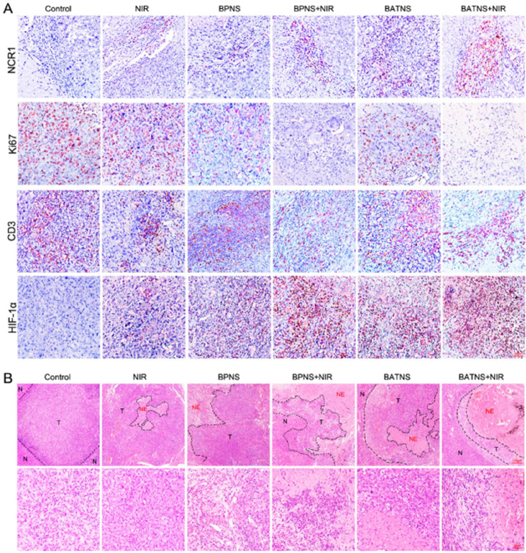 Figure 1
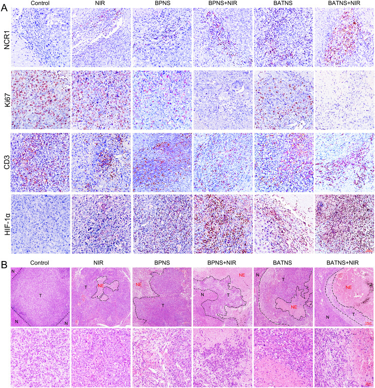 Figure 2
Peer Reviews
No public reviews on file for this paper yet. If you reviewed it on a platform where reviews are public (OpenReview, ICLR, NeurIPS, ICML), you can paste yours below so the community can read it here.
Videos
No videos yet. Explain this paper in a talk, walkthrough, or lecture? Add one.
Taxonomy
TopicsNanoplatforms for cancer theranostics · Extracellular vesicles in disease · Advanced Nanomaterials in Catalysis
Correction: Journal of Nanobiotechnology (2022) 20:90.
10.1186/s12951-022-01286-z
In this article, Fig. 7 appeared incorrectly and has now been corrected in the original publication. For completeness and transparency, the incorrect and correct versions of Fig. 7 are displayed below.
Incorrect Fig. 7.
Fig. 7. Analyze the changes of tumor histological indicators after BATNS photothermal treatment. A Immunohistochemical analysis of NK cells (NCR1), cell proliferation indicators (Ki67), T cells (CD3) and cellular hypoxia indicators (HIF-1α). B HE staining showed the local conditions of tumor tissue (T) adjacent to the cancer and necrotic tissue (NE) in the tumor in each experimental group. The tissue structure was observed under a ×100microscope (up), and the bottom under a ×400 microscope
Correct Fig. 7.
Fig. 7. Analyze the changes of tumor histological indicators after BATNS photothermal treatment. A Immunohistochemical analysis of NK cells (NCR1), cell proliferation indicators (Ki67), T cells (CD3) and cellular hypoxia indicators (HIF-1α). B HE staining showed the local conditions of tumor tissue (T) adjacent to the cancer and necrotic tissue (NE) in the tumor in each experimental group. The tissue structure was observed under a ×100microscope (up), and the bottom under a ×400 microscope
The original article has been corrected.