Endovascular treatment in comatose patients with anterior circulation ischemic stroke
Wouter M. Sluis, Simone M. Uniken Venema, Anouk van der Hoorn, Joseph C. J. Bot, Wim H. van Zwam, Jeannette Hofmeijer, H. Bart van der Worp

TL;DR
This study examines why some patients with a type of stroke become comatose and how they fare after treatment.
Contribution
The study identifies causes and outcomes of coma in a rare subgroup of stroke patients treated with endovascular thrombectomy.
Findings
Coma occurred in 1% of patients with anterior circulation ischemic stroke.
Comatose patients had worse outcomes and higher mortality rates.
Common causes of coma included bilateral ischemia and post-ictal states.
Abstract
Coma in the first hours after anterior circulation ischemic stroke is rare. We aimed to assess the causes of coma and outcomes after endovascular thrombectomy (EVT) in this relatively unexplored subgroup of patients. We used data from the MR CLEAN Registry, a prospective, multicenter, observational cohort study of patients treated with EVT in the Netherlands between March 2014, and December 2018. We included patients with anterior circulation ischemic stroke treated within 6.5 h of symptom onset and assessed frequency and causes of coma, defined as a score of 8 or lower on the Glasgow Coma Scale. Patients with a posterior circulation stroke were excluded. The primary outcome was the score on the modified Rankin Scale at 90 days. We compared outcomes of comatose and non-comatose patients with logistic regression. Fifty-two (1%) of 4,869 patients were comatose. The main causes of coma…
Genes, proteins, chemicals, diseases, species, mutations and cell lines named across the full text — each resolved to its canonical identifier and authoritative record.
Click any figure to enlarge with its caption.
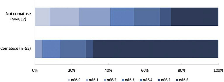 Figure 1
| Characteristics* | Not comatose | Comatose | |
|---|---|---|---|
| Clinical characteristics | |||
| Age, years (median; IQR) | 72.0 (62.0–81.0) | 73.5 (61.5–82.0) | |
| Sex (female) | 2,287 (47.5) | 31 (59.6) | |
| Pre-stroke functional dependency † | 600 (12.8) | 16 (32.7) |
|
| NIHSS (median; IQR) | 15.0 (11.0–19.0) | 25.0 (20.8–30.0) |
|
| Left hemisphere | 2,356 (52.5) | 39 (75.0) |
|
| In-hospital stroke | 501 (10.4) | 10 (19.2) | |
| Treatment with IVT | 3,514 (73.3) | 28 (53.8) |
|
| Systolic BP (mean; SD) | 150.1 (25.5) | 145.9 (35.3) | |
| Laboratory characteristics | |||
| INR (median: IQR) | 1.0 (1.0–1.1) | 1.1 (1.0–1.3) |
|
| CRP, mg/L (median; IQR) | 4.9 (2.0–11.0) | 4.0 (2.0–17.4) | |
| Glucose, mmol/L (median; IQR) | 6.8 (5.9–8.1) | 8.0 (6.6–10.3) |
|
| Imaging characteristics | |||
| ICA-T occlusion | 888 (19.2) | 20 (39.2) |
|
| ASPECTS |
| ||
| 0–4 | 193 (4.1) | 7 (13.7) | |
| 5–7 | 885 (19.0) | 10 (19.6) | |
| 8–10 | 3,590 (76.9) | 34 (66.7) | |
| Collateral score‡ |
| ||
| 0 | 244 (5.4) | 9 (18.0) | |
| 1 | 1,653 (36.4) | 18 (36.0) | |
| 2 | 1756 (38.6) | 18 (36.0) | |
| 3 | 891 (19.6) | 5 (10.0) | |
| Medical history | |||
| Hypertension | 2,513 (53.2) | 27 (52.9) | |
| Atrial fibrillation | 1,145 (24.1) | 16 (30.8) | |
| Diabetes | 809 (16.9) | 10 (19.2) | |
| Hypercholesterolemia | 1,448 (31.3) | 14 (27.5) | |
| Ischemic stroke | 850 (17.8) | 16 (30.8) |
|
| Myocardial infarction | 689 (14.6) | 7 (13.5) | |
| Peripheral arterial disease | 433 (9.2) | 5 (9.6) | |
| Medication use | |||
| Antiplatelet therapy | 1,495 (31.4) | 20 (40.0) | |
| DOAC | 227 (4.7) | 6 (12.0) |
|
| Coumarins | 603 (12.6) | 10 (19.6) | |
| Heparin | 146 (3.1) | 6 (12.0) |
|
| Statins | 1706 (36.2) | 20 (41.7) | |
| Anti-hypertensive agents | 2,623 (55.5) | 28 (56.0) | |
| Not comatose | Comatose | ||
|---|---|---|---|
| Treatment characteristics* | |||
| Onset to groin (mean; SD)† | 199.0 (74.4) | 225.3 (78.0) |
|
| Duration of procedure (mean; SD)† | 62.2 (34.4) | 65.8 (40.1) | |
| Onset to end of procedure (mean; SD)† | 253.2 (81.8) | 282.0 (78.3) |
|
| General anesthesia | 1,057 (23.3) | 30 (61.2) |
|
| Successful recanalization (TICI 2b-3) | 2,976 (64.4) | 39 (78.0) | |
| NIHSS after treatment (median; IQR)‡ | 9.0 (3.0–16.0) | 21.0 (15.0–25.0) |
|
| Not comatose | Comatose | ||
|---|---|---|---|
| Pneumonia* | 491 (10.2) | 6 (11.5) | |
| Intracranial hemorrhage | 278 (5.8) | 6 (11.5) | |
| Recurrent ischemic stroke | 73 (1.5) | 2 (3.8) | |
| Stroke progression | 416 (8.6) | 10 (19.2) |
|
| Not comatose | Comatose | aOR (95%CI) | |
|---|---|---|---|
| mRS at 90 days (median; IQR) | 3.0 (2.0–6.0) | 6.0 (4.0–6.0) | 6.11 (3.42–10.90)†† |
| 90-day mortality | 1,230 (26.4) | 35 (68.6) | 5.77 (3.22–10.34) |
| Futile recanalization† | 1,473 (32.6) | 36 (73.5) | 1.24 (0.79–1.95) |
Peer Reviews
No public reviews on file for this paper yet. If you reviewed it on a platform where reviews are public (OpenReview, ICLR, NeurIPS, ICML), you can paste yours below so the community can read it here.
Videos
No videos yet. Explain this paper in a talk, walkthrough, or lecture? Add one.
Taxonomy
TopicsAcute Ischemic Stroke Management · Traumatic Brain Injury and Neurovascular Disturbances · Stroke Rehabilitation and Recovery
Introduction
About one fifth of patients with acute ischemic stroke caused by a proximal arterial occlusion in the anterior circulation has a reduced consciousness on admission in the first 6 h of stroke onset (1), but coma, defined as a score on the Glasgow Coma Scale (GCS) of 8 or lower (2), is rare (3). In the first hours of anterior circulation ischemic stroke, coma may be caused by simultaneous involvement of both hemispheres, or by new ischemia in the hemisphere contralateral to a large previous hemispheric lesion. In about one third of the patients no underlying cause is found (3, 4). Coma could also be caused by stroke-induced epileptic seizures or by other causes than the stroke itself, such as hypoglycaemia or severe arterial hypotension (5).
Coma is not a contraindication to endovascular thrombectomy (EVT) in patients with anterior circulation ischemic stroke due to a large vessel occlusion (LVO) (6, 7), but the frequency of this condition and the outcomes after EVT are unknown. We therefore aimed to assess the frequency of coma in patients with ischemic stroke caused by a proximal occlusion in the anterior circulation, the causes of such coma, and the outcomes after EVT.
Materials and methods
Study protocol and population
We used data from the Multicenter Randomized Controlled Trial of Endovascular Treatment for Acute Ischemic Stroke in the Netherlands (MR CLEAN) Registry - a prospective, multicenter, observational study of patients with acute ischemic stroke treated with EVT in routine clinical practice in the Netherlands in 17 centers (8). The current study is based on data from patients included in the Registry between March 16, 2014, and December 31, 2018. Inclusion criteria for the present analysis were: age ≥ 18 years; a clinical diagnosis of acute ischemic stroke with a proximal arterial occlusion in the anterior circulation (internal carotid artery, middle cerebral artery [M1 and M2], or anterior cerebral artery [A1 and A2]), demonstrated with computed tomography angiography, magnetic resonance angiography, or digital subtraction angiography; and EVT initiated within 6.5 h of symptom onset or last seen well. We adhered to the RECORD guidelines (9). The checklist is attached as a Supplementary Table S1. The study protocol of the MR CLEAN Registry was reviewed by the Erasmus University Medical Center Ethics Committee, which served as the central review board for all participating centers. The requirement for written informed consent was waived. Patients or their representatives were provided with information on the study orally and in writing and were given the opportunity to refuse participation.
Data collection
Local investigators collected clinical and demographic data from patient records and assessed the score on the modified Rankin Scale (mRS) at 90 days (+/− 14 days).
Baseline imaging characteristics, including occlusion site, the Alberta Stroke Program Early CT Score (ASPECTS), and collateral status (10, 11), the extended treatment in cerebral infarction (eTICI) score after EVT (12), and follow-up imaging were assessed by the MR CLEAN Registry imaging core laboratory, blinded to clinical findings. Complications after treatment were assessed by a central serious adverse events adjudication committee, and included recurrent ischemic stroke, pneumonia, symptomatic intracranial hemorrhage (sICH, defined according to the Heidelberg Bleeding Classification) (13), and stroke progression (defined as neurological deterioration with an increase of ≥4 points on the NIHSS and follow-up imaging compatible with ischemia in the same territory without another underlying cause of deterioration). If no follow-up imaging was performed this was defined as neurological deterioration with an unknown cause.
As the GCS was not routinely collected in the Registry, written clinical information was evaluated for the GCS for all patients with a baseline score of 2 or higher on the level of consciousness question (item 1a) of the NIHSS. Patients were classified as comatose if the score on GCS derived from the written clinical information was 8 or lower. When patients were intubated and sedated for another reason (e.g., respiratory insufficiency due to a non-neurological cause), they were not counted as comatose if the GCS before intubation was known and above 8. If a pre-intubation GCS was not available, these patients were not considered comatose. The written clinical information of comatose patients was used to determine causes of coma.
Outcomes
The primary outcome measure was the score on the mRS at 90 (± 14) days. Secondary outcome measures were mortality at 90 days and futile recanalization, defined as functional dependence or death (mRS score, ≥ 3) at 90 days despite successful recanalization (eTICI ≥2b) at the end of EVT.
Statistical analysis
The frequency and causes of coma are described with descriptive statistics. Comparisons between baseline characteristics of patients who were comatose and those who were not were made with χ2, Student’s t or Mann–Whitney U test, where appropriate. We used multivariable (ordinal) logistic regression to calculate adjusted (common) odds ratios (a(c)OR) for all outcomes and adjusted for age, pre-stroke score on the mRS, NIHSS at baseline, onset-to-groin time, history of diabetes, history of atrial fibrillation, and treatment with intravenous thrombolysis. For the multivariable regression analyses, multiple imputation was performed for predictor values with missing data. A sensitivity analysis was performed without using multiple imputation for missing data. Statistical significance was defined as a p value <0.05. All statistical analyses were performed with R studio version 1.3.1056. (Rstudio PBC).
Results
A total of 4,869 patients were included in this analysis (Supplementary Figure S1). Fifty-two (1%) patients were comatose before EVT. In 20 patients (39%) the cause of coma was a radiologically apparent bilateral ischemic lesion. Twelve patients had acute ischemic stroke in the presence of a previous cortical ischemic stroke in the contralateral hemisphere and eight had acute bilateral ischemic stroke, of whom six had a bilateral large vessel occlusion (LVO). Only one patient with a bilateral LVO was treated with EVT on both sides, which was only successful for one side. Of the remaining comatose patients, nine (17%) were respiratory insufficient, five (10%) were in a post-ictal state after a seizure, two (4%) were hypotensive due to cardiogenic shock, and the other patients were either hypothermic (1 patient, 2%), intoxicated with alcohol (1 patient, 2%), post-anoxic after cardiac arrest (1 patient, 2%) or in septic shock (1 patient, 2%). In twelve patients (23%) no clear cause of coma could be determined. Six of these patients eventually died of space-occupying edema, which was not visible on non-contrast CT before EVT. None of these patients was treated with decompressive surgery.
Patients who were comatose were comparable to non-comatose patients with respect to age, sex, medication, and comorbidities but were more frequently functionally dependent before the stroke (33% vs. 13%, p < 0.001), had a higher NIHSS (25 vs. 15, p < 0.001), more often a stroke of the left hemisphere (75% vs. 53%, p = 0.002), or a carotid-T occlusion [internal carotid artery (ICA) + middle cerebral artery (MCA), 39% vs. 19% (p = 0.001)]. Fewer comatose patients were given IVT (54% vs. 73%, p = 0.003) and they had worse ASPECTS and collateral scores (Table 1). Two comatose patients (3.8%) had a very low ASPECTS score (0 through 2), which was slightly higher than the group of patients without coma (n = 51, 1.1%).
Onset-to-groin times were longer for comatose patients (225 vs. 199 min, p = 0.012) and treatment was more often performed under general anesthesia (61% vs. 23% p < 0.001). The NIHSS at 24 to 48 h was higher in the comatose group (21 vs. 9, p < 0.001) and comatose patients were more often diagnosed with stroke progression (19% vs. 9%, p = 0.015) (Tables 2, 3).
After adjustment for confounders, comatose patients died more often within 90 days (69% vs. 26%; aOR 2.95; 95%CI:1.47–5.90; Tables 1, 4; Figure 1), with a median time to death of 3 days (IQR 2–7).
90-day mRS of patients with or without coma. mRS = modified Rankin Scale.
Mortality was highest in comatose patients with a bilateral ischemic lesion (70%) or a non-neurological cause of coma (71%) and lowest in the post-ictal patients (40%). Comatose patients had a worse functional outcome at 90 days (acOR for a shift on the mRS towards a worse functional outcome: 2.73; 95%CI: 1.45–5.13; Table 4; Figure 1) and just two patients (4%) were functionally independent at 90 days: one patient in a post-ictal state after a seizure and one with an unknown cause of coma. Recanalization was futile in 74% of comatose patients. The sensitivity analysis without imputed data yielded similar results (Supplementary Table S2).
Discussion
In this study, patients with ischemic stroke caused by a proximal occlusion in the anterior circulation who were comatose within 6.5 h of stroke onset had a very high risk of death or dependency at 90 days after EVT, even with angiographic recanalization. The most frequent cause of coma was an ischemic lesion in the hemisphere contralateral to the occlusion.
Acute bilateral large vessel occlusion occurs in less than 1% of patients with ischemic stroke. The prognosis is generally poor, as was the case in our six patients (4). For unknown reasons, only one of them was treated on both sides and recanalization only occurred on one side. We therefore do not know whether outcomes would have been better if patients with bilateral occlusions had bilateral recanalization. Contralateral recurrent stroke occurred in 7% of patients in The Copenhagen Stroke Study and was also associated with a worse functional outcome compared to ipsilateral recurrence (14).
Most patients with a systemic non-neurological cause of coma had signs of severe metabolic, respiratory or hemodynamic dysregulation. Hypotension, hypoxia, and infections after stroke have been associated with a worse prognosis (15–17). In about one quarter of comatose patients, no cause of reduced consciousness was identified, which is comparable to a previous study in which no cause of coma was found (3). Remarkably, in our study more than half of the patients with coma of unidentified cause died of space-occupying edema in the next days, without signs of mass effect on non-contrast CT before EVT, none of the patients in our study were treated with decompressive surgery, as opposed to around 40% in a previous MR CLEAN study (18). With the exception of one patient, all these patients primarily presented to the intervention center, so most imaging was directly prior to EVT. Space-occupying edema is a well-known cause of coma in patients with unilateral anterior circulation stroke, but clear clinical signs of mass effect generally occur after the first few hours, and only in one third of the cases within 24 h (19, 20). In a study of patients with middle cerebral artery infarction caused by an M1 occlusion, 15 of the 24 patients (63%) who eventually developed ‘malignant’ space-occupying edema had a reduced level of consciousness in the first 6 hours of stroke onset, but the proportion of patients with coma in this study is not known (21).
The finding that comatose patients were less frequently treated with IVT and had longer door-to-groin times could suggest that treating physicians are hesitant to treat comatose patients, possibly due to a lack of evidence on the efficacy of reperfusion therapy in this subgroup of patients. Another explanation could be a greater diagnostic delay, and therefore missing the time window for treatment with IVT, or suspected comorbidity with a suspected increased risk hemorrhage. In a study on IVT in patients with ischemic stroke and a decreased consciousness, IVT seemed beneficial, but this study was just as the present study not randomized and retrospective in design, possibly causing selection bias (22).
The current study is the first to report causes of coma and outcomes of EVT in patients with anterior circulation ischemic stroke, but there are limitations to consider. First, this is a retrospective analysis in a prospective registry of patients who were treated with EVT. We therefore cannot provide information on comatose patients with anterior circulation ischemic stroke who were not treated with EVT, for example because they were considered by the clinician as too severe for curative treatment. Second, causes of coma were determined retrospectively based on written clinical information, which may be insufficient to accurately reflect the actual causes of coma. EEG was not routinely performed and the presence or absence of a fetal posterior communicating artery was not assessed. Third, the group of patients with coma was very small compared to the non-comatose group and therefore the uncertainty of our estimates is considerable. Fourth, patients included in the MR CLEAN Registry were treated early in the era of EVT. Since then, devices and skills have improved which could positively influence outcomes of comatose patients. Nevertheless, our data strongly suggest that the risk of a poor outcome after EVT is high in patients who are comatose on admission is, especially in patients with bilateral ischemic stroke. Further research is needed to assess the benefit of EVT in patients without a known cause of coma.
Conclusion
Patients with anterior circulation ischemic stroke who are comatose before endovascular thrombectomy in the first 6.5 h often have ischemic lesions in both hemispheres or an underlying metabolic or hemodynamic dysregulation as the cause of their decreased consciousness. These patients have a very poor prognosis despite successful EVT.
The reference list from the paper itself. Each links out to its DOI / PubMed record.
- 1Janssen PM Chalos Vvan den Berg SA Kompanje EJO Nederkoorn P Jvan der Worp BH. Neurological deficits in stroke patients that may impede the capacity to provide informed consent for endovascular treatment trials. J Stroke Cerebrovasc Dis. (2019) 28:104447. doi: 10.1016/j.jstrokecerebrovasdis.2019.10444731624035 · doi ↗ · pubmed ↗
- 2Jennett B Teasdale G. Aspects of coma after severe head injury. Lancet. (1977) 1:878–81. doi: 10.1016/S 0140-6736(77)91201-6, PMID: 67287 · doi ↗ · pubmed ↗
- 3Young MJ Awad A Andreev A Bonkhoff AK Schirmer MD Dmytriw AA. Characterizing coma in large vessel occlusion stroke. J Neurol. (2024) 271:2658–61. doi: 10.1007/s 00415-024-12199-2, PMID: 38366071 PMC 11889655 · doi ↗ · pubmed ↗
- 4Kwon SU Lee SH Kim JS. Sudden coma from acute bilateral internal carotid artery territory infarction. Neurology. (2002) 58:1846–9. doi: 10.1212/WNL.58.12.1846, PMID: 12084889 · doi ↗ · pubmed ↗
- 5Horsting MW Franken MD Meulenbelt Jvan Klei W Ade Lange DW. The etiology and outcome of non-traumatic coma in critical care: a systematic review. BMC Anesthesiol. (2015) 15:65. doi: 10.1186/s 12871-015-0041-9, PMID: 25924678 PMC 4424591 · doi ↗ · pubmed ↗
- 6Turc G Bhogal P Fischer U Khatri P Lobotesis K Mazighi M. European stroke organisation (ESO) - European Society for Minimally Invasive Neurological Therapy (ESMINT) guidelines on mechanical Thrombectomy in acute Ischaemic Stroke Endorsed by stroke Alliance for Europe (SAFE). Eur Stroke J. (2019) 4:6–12. doi: 10.1177/2396987319832140, PMID: 31165090 PMC 6533858 · doi ↗ · pubmed ↗
- 7Powers WJ Rabinstein AA Ackerson T Adeoye OM Bambakidis NC Becker K. Guidelines for the early Management of Patients with Acute Ischemic Stroke: 2019 update to the 2018 guidelines for the early Management of Acute Ischemic Stroke: a guideline for healthcare professionals from the American Heart Association/American Stroke Association. Stroke. (2019) 50:e 344–418. doi: 10.1161/STR.000000000000021131662037 · doi ↗ · pubmed ↗
- 8Jansen IGH Mulder MJHL Goldhoorn RB. MR CLEAN registry investigators. Endovascular treatment for acute ischaemic stroke in routine clinical practice: prospective, observational cohort study (MR CLEAN registry). BMJ. (2018) 360:k 949. doi: 10.1136/bmj.k 949, PMID: 29523557 PMC 5844245 · doi ↗ · pubmed ↗