Spinal Phlegmon Secondary to Neisseria meningitidis Meningitis With Possible Herpes Simplex Virus 1 Co-infection in an Immunocompetent Pediatric Patient
Peter Paul Lim, Kayla Morisette, Meredith Keith, Eric Hines, Mark K Huntington

TL;DR
A 5-year-old child with meningitis caused by Neisseria meningitidis and possibly HSV-1 developed a rare spinal infection, with a successful recovery after treatment.
Contribution
First reported case of spinal phlegmon with meningococcal meningitis and HSV-1 co-infection in an immunocompetent child.
Findings
Spinal phlegmon occurred in a child with meningococcal meningitis and HSV-1 co-infection.
The patient showed no signs of immunodeficiency despite the rare polymicrobial infection.
Treatment with prolonged antibiotics and antivirals led to a favorable outcome.
Abstract
Spinal phlegmon has rarely been reported in meningococcemia. Simultaneous polymicrobial infection has been occasionally reported as the etiology of meningitis. The development of a spinal phlegmon and optic neuritis complicated meningitis in a five-year-old male who presented with clinical sepsis and meningitis. Neisseria meningitidis (N. meningitidis) and herpes simplex virus 1 (HSV-1) were demonstrated in the cerebrospinal fluid. Investigation of potential immunocompromise revealed no identified deficit. Protracted antibiotics and antiviral therapy were completed with a favorable clinical outcome. This is potentially the first reported case of spinal phlegmon associated with meningococcal meningitis in an immunocompetent pediatric patient. It occurred in the clinical context of what appeared to be a simultaneous infection by N. meningitidis and HSV-1.
Genes, proteins, chemicals, diseases, species, mutations and cell lines named across the full text — each resolved to its canonical identifier and authoritative record.
Click any figure to enlarge with its caption.
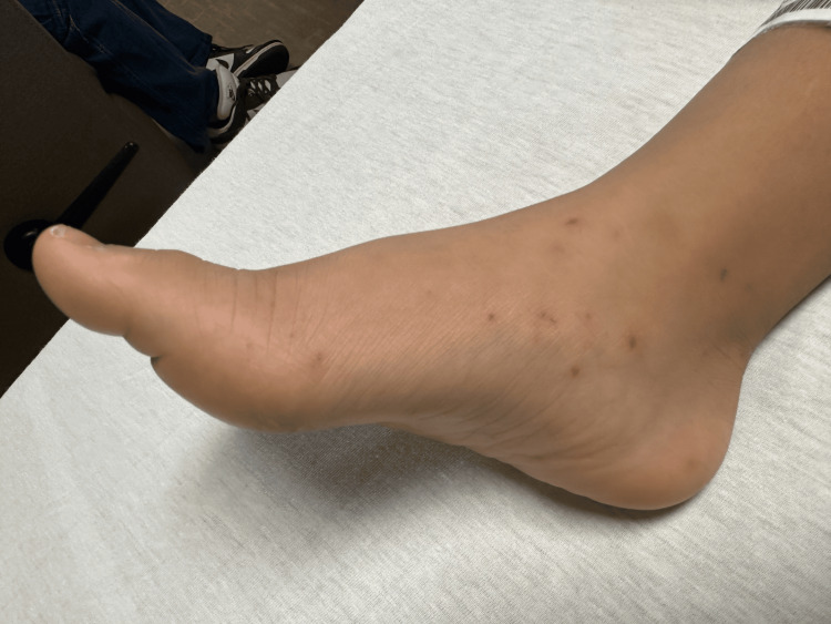 Figure 1
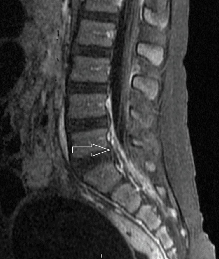 Figure 2
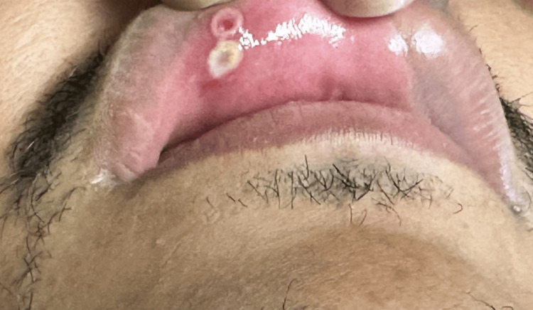 Figure 3
| Hospital day collected | Source | Test | Results (normal) | Comments |
| 1 | CSF | Erythrocyte count (RBC) | 5750/uL | Fluid was yellow and cloudy |
| Nucleated cells | 13500/uL (<7) | |||
| 96% neutrophils | ||||
| 4% lymphocytes | ||||
| Glucose | <10 mg/dL (40-70) | |||
| Total protein | 421 mg/dL (40-70) | |||
| Gram stain | Gram negative diplococci | |||
| PCR | Positive for | |||
| Positive for HSV-1 | ||||
| Culture | No growth* | |||
| Blood | Leukocyte count (WBC) | 8.5 K/uL (5-15.5) | - | |
| 91.6% neutrophils (38.2-45.8) | ||||
| 4.1% lymphocytes (45.5-54.5) | ||||
| 3.3% monocytes (4.5-5.5) | ||||
| 0% eosinophils (2.7-3.3) | ||||
| Hemoglobin | 13.3 g/dL (11.5-13.5) | |||
| Platelets | 146K/uL (150-350) | |||
| Sodium | 139 mmol/L (135-147) | |||
| Potassium | 2.8 mmol/L (3.4-4.7) | |||
| Chloride | 106 mmol/L (97-107) | |||
| Bicarbonate | 19 mmol/L (17-26) | |||
| BUN | 18 mg/dL (9-22) | |||
| Creatinine | 0.5 mg/dL (0.3-0.6) | |||
| ALT | 12 U/L (9-23) | |||
| AST | 20 U/L (20-40) | |||
| ALP | 224 U/L (132-315) | |||
| CRP | 50.8 mg/dL (<0.5) | |||
| Total bilirubin | 0.9 ng/dL (0.1-0.4) | |||
| total protein | 7.5 g/dL (5.9-7.2 | |||
| Blood culture | No growth* | |||
| Imaging | CT of the head | No pathology | ||
| MRI of the head | No pathology | |||
| MRA of the head | No pathology | |||
| MRV of the head | No pathology | |||
| 5 | CSF | Erythrocyte count (RBC) | 4643 /uL | Fluid was colorless and cloudy |
| Nucleated cells | 1217 /uL (<7) | |||
| 5% neutrophils | ||||
| 95% lymphocytes | ||||
| Glucose | 31 mg/dL (40-70) | |||
| Total protein | 102 mg/dL (40-70) | |||
| Gram stain | negative | |||
| PCR** | Positive for | |||
| Negative for HSV-1 | ||||
| Positive for HHV-6 | ||||
| Viral culture | No growth | Collected post-antiviral treatment | ||
| 7 | Imaging | MRI of the brain | Small focus of enhancement within or adjacent to the apex of the occipital horn of the right lateral ventricle, possibly involving the ependymal | - |
| MRI of the spine (C, T, L) | Enhancement of nerve roots of the cauda equina. Prominent hyperenhancement in the ventral epidural space at the L5-S2 levels, concerning for developing phlegmon or early abscess | |||
| Outpatient follow-up | Blood | Complement levels | Normal | Complement deficiency is associated with increased risk for meningococcal infection |
| Genetic testing | Primary immunodeficiency panel | No deficiencies identified | - | |
| Imaging | MRI of the brain | Persistent focal ependymal/subependymal enhancement of the right occipital horn | - | |
| MRI of the spine | Persistent inflammation of cauda equina. Prior epidural space finding resolved | - |
| Coinfection | Organism |
| Bacterial | Neisseria + Bordetella |
| Neisseria + Streptococcus | |
| Viral |
|
| Fungal | Neisseria + Cryptococcus |
Peer Reviews
No public reviews on file for this paper yet. If you reviewed it on a platform where reviews are public (OpenReview, ICLR, NeurIPS, ICML), you can paste yours below so the community can read it here.
Videos
No videos yet. Explain this paper in a talk, walkthrough, or lecture? Add one.
Taxonomy
TopicsBacterial Infections and Vaccines · Infectious Diseases and Tuberculosis · Amoebic Infections and Treatments
Introduction
Meningitis is an inflammation of the lining surrounding the brain and spinal cord due to an infectious process. While fungal and parasitic causes exist, viruses and bacteria are the main pathogens. The most common etiology is viral infection; the most severe is bacterial infection [1].
A wide range of viruses may cause meningitis, including herpesviruses, enteroviruses, adenoviruses, arboviruses, lymphocytic choriomeningitis virus, and mumps virus. For a majority of these, treatment is primarily supportive, though antiviral drugs have an important role in some, especially in herpesvirus infection.
Bacterial causes of meningitis include the genera of Enterobacter, Escherichia, Haemophilus, Listeria, Mycobacterium, Neisseria, Orientia, Pseudomonas, Salmonella, Staphylococci, and Streptococci. These may be rapidly progressive, complicated by abscess formation, and may lead to sepsis. Early diagnosis and antibiotic treatment are critical.
Herpes simplex virus (HSV) is one of the most common viral etiologies of meningitis; type 2 is more commonly associated with meningitis than type 1. Transmission is generally via mucosal contact (genital or oral). Long-term neurological sequelae are more common with HSV than in other viral meningitis cases, and it may cause recurrent aseptic meningitis (Mollaret’s meningitis). Early diagnosis and antiviral treatment greatly improve outcomes [2,3].
Neisseria meningitidis (N. meningitidis), also known as meningococcus, is a Gram-negative diplococcus that causes severe meningitis and septicemia. It may progress rapidly to death. It is spread by exposure to respiratory and throat secretions, including those of asymptomatic carriers. Age groups with higher rates of meningococcal disease include infants in their first year of life, those ages 16-23 years old, and those over age 85 years old. Individuals with asplenia, human immunodeficiency virus (HIV) infection, or complement deficiencies or those on complement pathway inhibitors are significantly at risk for infection [1].
Meningitis occurring coincident with other systemic infections by a different organism is not rare, but coinfection of the cerebrospinal fluid (CSF) by multiple pathogens as a cause of meningitis is uncommon. These polymicrobial infections are usually associated with neurosurgical procedures, but community-acquired cases, including bacterial-viral coinfections, have been reported [4]. Coinfection with Neisseria and HSV has not been previously reported.
We present a case of meningitis with phlegmon attributable to coinfection by N. meningitidis and HSV.
Case presentation
A five-year-old male with a history of speech delay presented to a stand-alone emergency department with an approximately four-day history of decreased energy, non-bilious, non-bloody emesis, loose stools, and fevers followed by acute development of rash in the back and lower extremities, along with subsequent confusion and somnolence.
On initial examination, his vital signs showed a temperature of 98°F, pulse 101, respiratory rate 20, blood pressure 105/70 mmHg, and room air oxygen saturations of 100%. He was irritable, ill-appearing, and lethargic, with altered mental status and difficulty following commands or responding to questions. Cranial nerves II-XII were intact, pupils sluggish in response to light, no papilledema. There was subtle, non-rigid flexion of the upper extremities and extension of the lower extremities. The deep tendon reflexes were intact. Several petechial to purpuric lesions were noted on his face, back, and lower and upper extremities with involvement of the soles of his feet (Figure 1). He was also noted to have nuchal rigidity. Laboratory evaluation showed a normal leukocyte (WBC) count but predominance of segmented leukocytes, thrombocytopenia, hypokalemia, hyperbilirubinemia, and significantly elevated inflammatory markers (Table 1).
Purpuric rash
Empiric meningitic vancomycin dosing and ceftriaxone were started immediately; blood and CSF samples were obtained following antibiotic administration. Dexamethasone was also started around the same time antibiotics were initiated. Emergent computed tomography of the brain with contrast was done prior to lumbar puncture (LP) to rule out contraindication to CSF sampling, which was read as reassuring (Table 1). An LP was immediately done, and CSF output was noted to be cloudy. CSF microscopic analysis revealed significant pleocytosis of 13,500 total nucleated cells (normal (N) 0-7 cells/uL) with 96% neutrophils, hypoglycorrhachia of <10 mg/dL (N 40-70 mg/dL), total protein of 421 mg/dL (N 15-45 mg/dL), and erythrocytes (RBC) of 5750 cells/uL (N 0 cell/uL). A Gram stain of the CSF showed Gram-negative diplococci. Molecular testing of the CSF was positive both for N. meningitidis and HSV-1. Acyclovir was immediately added to his regimen.
The patient’s clinical course worsened acutely on the day of admission, with a deteriorating neurologic status that prompted extensive imaging investigation with magnetic resonance imaging (MRI) with and without contrast enhancement, magnetic resonance angiography, and magnetic resonance venography of the brain to rule out increased neurosurgical emergency. All of these baseline studies were reported as normal.
The patient’s sentinel CSF culture demonstrated growth of N. meningitidis. Repeat LP was performed on day 5 of hospitalization to reassess CSF parameters and to determine CSF microbiologic clearance. Subsequent CSF examination showed total nucleated cells of 1217 with a 95% lymphocytic predominance; polymerase chain reaction (PCR) remained positive for N. meningitidis, was positive for human herpesvirus 6 (HHV-6), and negative for HSV-1 (Table 1).
While the patient demonstrated overall clinical improvement by day 5, gait instability and intermittent fevers to 104.6°F persisted. Brain MRIs and MRIs of the cervical, thoracic, and lumbar spine were obtained after 7 days of hospitalization. The brain MRI showed a new 5 mm focus of hyperintense signal on diffusion either within the atrium of the right lateral ventricle or possibly involving the ependymal surface. There was a subtle focus of enhancement at the apex of the occipital horn of the right lateral ventricle involving the ependyma. This may represent a small purulent material or a microinfarct secondary to meningitis. The cervical spine MRI was negative for findings. The thoracic and lumbar spine MRI showed enhancement of nerve roots of the cauda equina consistent with the patient’s history of meningitis, mild prominence of the upper thoracic region with subtle stranding enhancement representing inflammation or infection, and a prominent hyperenhancement in the L5-S2 level concerning for inflammation, infection, and possible early development of spinal phlegmon (Figure 2).
MRI showing prominent enhancement of the ventral epidural space of L5-S1 representing the developing phlegmon (arrow)MRI: magnetic resonance imaging
To evaluate whether the HSV-1 results truly represented a bona fide co-infection, repeat testing of the sentinel CSF sample was performed. Only N. meningitidis was positive on the multiplex PCR (BiofireTM, Salt Lake City, Utah) panel. A stand-alone HSV-1 PCR test was negative. Serum HSV 1 and 2 IgG was positive at 13.60, and HSV 1 and 2 IgM was elevated at 0.98 (ref: ≤0.89), suggesting HSV-1 was a bona fide active infection. The patient’s father was later found to have resolving mucosal ulcers in his upper lip that clinically looked like HSV gingivostomatitis. This was swabbed and negative for HSV-1 and HSV-2 by PCR; however, the specimens were obtained on day 11 of his course following a course of acyclovir, implicating theoretically decreased viral load from the sample and a suspected false negative result. The father’s lesions revealed that this has been recurrent, often painful, and most often incited by sunlight exposure, strongly suggesting occult HSV gingivostomatitis.
The patient completed a 21-day course of acyclovir 10 mg/kg every eight hours and a six-week total course of ceftriaxone 50 mg/kg every 12 hours with overall clinical improvement. His surveillance MRI prior to cessation of therapy showed significant resolution of spinal enhancement and overall radiologic findings. He underwent an extensive immune workup, which did not reveal any inborn immunity error or other immunological deficit. He was back to baseline neurologic function on his follow-up after completion of treatment. Hearing evaluation revealed no deficits, but unfortunately, his ophthalmologic exam showed optic neuritis as a complication of his meningoencephalitis that was stable on subsequent ophthalmologic follow-up and did not need additional ancillary intervention.
Discussion
N. meningitidis is a Gram-negative diplococcus responsible for meningococcal disease, ranging from meningitis to disseminated disease [5]. Mortality is 50%, 10-15% even with proper treatment. The development of the purpuric rash in our patient suggests that the presentation was in the early stages of a disseminated disease. Patients aged <1 year, 16-23 years, or >85 years are at greater risk for severe disease, as are those with asplenia, complement deficiency, or HIV [6]. Our patient did not fit into any of these demographics.
A novel aspect of this patient's course is the development of spinal myelitis and phlegmon, which are presumed to be caused by N. meningitidis infection. While spinal epidural abscess and myelitis have been reported as complications of Streptococcus pneumoniae and Escherichia coli, relevant literature is scarce on spinal cord complications of N. meningitidis infection [7-11]. Cerebral suppurative complications of N. meningitidis are very rare infectious phenomena. While N. meningitidis has been reported to produce abscesses in various locations, including the brain, we could not identify any previous reports of spinal abscess/phlegmon associated with the organism.
HSV can cause meningitis or encephalitis. HSV-1 is most commonly associated with encephalitis, while HSV-2 is often associated with meningitis and more often implicated in genital infection [12,13]. Central nervous system infections with HSV-1 are more deadly than those of HSV-2; for HSV-1 encephalomeningitis, the only modifiable risk factor that can affect outcomes is prompt initiation of intravenous acyclovir [3].
A recent study identified mixed bacterial-viral infections as the cause of meningitis in 0.9% of cases [4]. Meningitis due to both meningococcus and another pathogen has been previously reported with various organisms, including Cryptococcus, Bordetella, and Streptococcus, but not HSV (Table 2) [14-17].
The identification of meningococcus by Gram stain, culture, and PCR incontrovertibly establishes the presence of this agent and its role in our patient’s disease. Several factors support the legitimacy of the diagnosis of coinfection with HSV-1. The repeat LP showed a white count of 1200/µL, with 95% lymphocytes, strongly suggesting an ongoing active viral component of the infectious process. The cytology should have theoretically maintained a neutrophilic predominance if it were solely due to bacterial infection. Serological studies demonstrated an elevated HSV IgM, consistent with an acute infection. Following these results, further history attempting to identify a possible source revealed the patient’s father had developed cold sores the week prior to presentation (Figure 3).
Buccal HSV lesionsHSV: herpes simplex virus
Although the presence of the virus was not conclusively demonstrated (viral culture and viral load were not checked as they would add cost to care without altering management), we believe active HSV infection was present. Regardless of whether the HSV component was a reactivation or a new active infection, the sensitive detection by PCR in the CSF is an irrevocable finding that necessitates a cautious clinical approach. Although we cannot confirm this retrospectively, even the minute possibility that active infection was present dictates that clinical management should be sagacious in similar clinical settings. It is tempting to speculate that the unusual development of phlegmon in this case of meningococcus was facilitated by immunosuppression from HSV-1, which is believed to affect bacterial infections in adult intensive care unit patients [18].
A positive HHV-6 PCR on the second LP implicates the question of a triple co-infection, which may be very unlikely. HHV-6 can be chromosomally integrated and is positive in CSF for up to 17% of the population. Therefore, it often has inconsequential implications, especially among immunocompetent patients [19,20]. The HHV-6 was tested twice on hospital day 1 and was negative on both multiplex tests.
Exploration of possible immune compromise showed no evidence of primary immunodeficiency, and complement test results were normal (Table 1).
Conclusions
We report here what we believe to be potentially the first case in the English language literature of phlegmon in meningococcus meningitis. We also report a case of meningitis due to potential coinfection with HSV-1. With nearly 1% of meningitis cases being due to coinfection, this is a reminder of the importance of continued investigation for potential etiological agents even after one is identified, especially if the additional infectious agent identified might lead to a change in clinical management. The misapplication of Occam’s razor could result in missing potentially lethal contributors to the patient’s illness.
The reference list from the paper itself. Each links out to its DOI / PubMed record.
- 1Meningitis 8 2024 2023 https://www.who.int/news-room/fact-sheets/detail/meningitis
- 2Prevalence of herpes simplex virus infections in the central nervous system Clin Lab Akkaya O 67202110.7754/Clin.Lab.2020.20111134258967 · doi ↗ · pubmed ↗
- 3Viral meningitis and encephalitis: an update Curr Opin Infect Dis Gundamraj V Hasbun R 1771853620233709304210.1097/QCO.0000000000000922 · doi ↗ · pubmed ↗
- 4Dynamics of community-acquired meningitis syndrome outbreaks in southern France Front Microbiol Morsli M Salipante F Kerharo Q 11021301320223677702910.3389/fmicb.2022.1102130 PMC 9909019 · doi ↗ · pubmed ↗
- 5Clinical Overview of Meningococcal Disease 8 2024 2024 https://www.cdc.gov/meningococcal/hcp/clinical/index.html
- 6Demographic profiles and risk factors for mortality in acute meningitis: a nationwide population-based observational study Acute Med Surg Akaishi T Tarasawa K Fushimi K Yaegashi N Aoki M Fujimori K 92011202410.1002/ams 2.920PMC 1075699038162167 · doi ↗ · pubmed ↗
- 7Spinal and paraspinal pneumococcal infections-a review Eur J Clin Microbiol Infect Dis Siddiq DM Musher DM Darouiche RO 5175273320142448174810.1007/s 10096-013-1997-3 · doi ↗ · pubmed ↗
- 8A case report of multiple cerebral abscess formation complicating serogroup B Neisseria meningitidis meningitis BMC Infect Dis O'Connor C Collins A Twomey E Hensey C Caird J Gavin PJ 8631920193163891310.1186/s 12879-019-4509-y PMC 6802112 · doi ↗ · pubmed ↗