Leukocytoclastic Vasculitis: Distinguishing Drug-Induced From Hepatitis C-Related Causes
Charles J DeBiase, Stephen K Stacey, Richard J LaBaere

TL;DR
A 34-year-old woman with a complex medical history presented with symptoms suggesting vasculitis, highlighting the challenge of distinguishing between drug-induced and hepatitis C-related causes.
Contribution
The paper presents a case emphasizing the diagnostic challenge of differentiating drug-induced vasculitis from hepatitis C-related vasculitis.
Findings
The patient's symptoms were initially suggestive of hepatitis C-related mixed cryoglobulinemia.
Further evaluation suggested drug-induced vasculitis as the more likely cause.
The case underscores the importance of thorough evaluation for accurate diagnosis and management.
Abstract
This report presents the case of a 34-year-old female with chronic cholecystitis and hepatic failure due to alcohol use and hepatitis C. She also has a history of opioid and alcohol use disorders. Frequent hospitalizations for fluid overload culminated in her presentation with lower extremity swelling and palpable purpura. Investigation revealed leukocytoclastic vasculitis, initially raising concern for mixed cryoglobulinemia from hepatitis C but ultimately more likely due to drug-induced vasculitis. This case highlights the diagnostic challenge of differentiating between hepatitis C-associated mixed cryoglobulinemia and drug-induced vasculitis, emphasizing the need for thorough evaluation to guide appropriate management.
Genes, proteins, chemicals, diseases, species, mutations and cell lines named across the full text — each resolved to its canonical identifier and authoritative record.
Click any figure to enlarge with its caption.
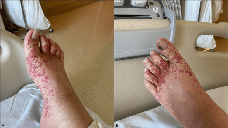 Figure 1
| Parameter | Normal range (units) | Patient results (on admission) |
| Hemoglobin | 11.0-14.5 g/dL | 8.3 |
| Platelets | 150-450 x 10⁹/L | 110 |
| AST | 0-33 U/L | 62 |
| Direct bilirubin | 0.1-0.3 mg/dL | 8.9 |
| ESR | 0-20 mm/hr | 12 |
| CRP | 0-10 mg/L | 6.4 |
| Lactate | 0.5-2.2 mmol/L | 2.1 |
| C4 | 10-40 mg/dL | 31 |
| HIV | Negative | Negative |
| Hepatitis C viral load | Undetectable | 1,040,000 |
| Rheumatoid factor | <14 IU/mL | Negative |
| Cryoglobulin serology | Negative | Negative |
| Serum protein electrophoresis | Normal | Normal |
Peer Reviews
No public reviews on file for this paper yet. If you reviewed it on a platform where reviews are public (OpenReview, ICLR, NeurIPS, ICML), you can paste yours below so the community can read it here.
Videos
No videos yet. Explain this paper in a talk, walkthrough, or lecture? Add one.
Taxonomy
TopicsHeparin-Induced Thrombocytopenia and Thrombosis · Hepatitis C virus research · Vasculitis and related conditions
Introduction
Leukocytoclastic vasculitis (LCV) is an immune-mediated small vessel vasculitis characterized by neutrophilic infiltration and fibrinoid necrosis of postcapillary venules, often presenting as palpable purpura on the lower extremities. It can be triggered by infections, autoimmune conditions, or medications [1]. The global incidence of cutaneous LCV varies from 15 to 38 cases per million per year, depending on the definition used. The etiology remains unidentified in approximately half of the cases [2]. In our patient, the potential causes included her chronic hepatitis C infection or recent antibiotic use due to the recent addition of metronidazole and cefdinir, both of which are known triggers for vasculitis. Other common causes of drug-induced vasculitis to consider include NSAIDs, diuretics, and antithyroid medications [1]. Distinguishing between underlying etiologies is critical, as treatment varies depending on the cause. Here, we present a case of a 34-year-old female with chronic hepatic failure from alcohol use and hepatitis C who developed lower extremity palpable purpura, initially raising concern for cryoglobulinemia-associated vasculitis but ultimately determined to be likely drug-induced. This case underscores the need for careful evaluation in patients with vasculitic presentations to ensure accurate diagnosis and appropriate management.
Case presentation
A 34-year-old female with hepatic failure secondary to alcohol use (currently in remission), untreated hepatitis C, and opioid use disorder presented to the emergency department with lower extremity swelling and palpable purpura that had begun earlier that day. She had been discharged the previous day after treatment for acute-on-chronic cholecystitis and was prescribed oral cefdinir and metronidazole upon discharge for continued antimicrobial coverage targeting biliary pathogens.
On presentation, she reported ongoing right upper quadrant pain rated as a 7/10, consistent with her level of pain at discharge from the previous hospital stay. She denied shortness of breath, chest pain, fevers, chills, and nausea. Examination revealed jaundice and tender, palpable purpura clustered on her trunk and extremities, being the most severe on the dorsal feet (Figure 1). Right upper quadrant tenderness was also noted, consistent with her discharge examination. No other significant findings were observed.
Images of the patient's right (A) and left (B) feet showing multiple palpable purpuric lesions, predominantly clustered on the dorsal surfaces.
In the emergency department, she had a temperature of 36.2°C, blood pressure of 137/75 mmHg, heart rate of 105 beats per minute, respirations of 18 breaths per minute, and oxygen saturation of 97%. Complete blood count, comprehensive metabolic panel, and labs were obtained to evaluate the likely etiology of her palpable purpuric lesions (Table 1). Findings were notable for anemia (hemoglobin 8.30 g/dL), thrombocytopenia (110,000 platelets per microliter), mildly elevated aspartate aminotransferase (62 U/L), and elevated direct bilirubin (8.9 mg/dL). Hepatitis C viral load was high (1,040,000 international units per milliliter). C-reactive protein, lactate, and complement levels were within normal limits. HIV, rheumatoid factor, and cryoglobulin serology testing were negative. Serum protein electrophoresis was negative. A biopsy of the lesions on her foot showed LCV.
Based on the patient’s palpable purpura and skin biopsy findings, the differential diagnosis included mixed cryoglobulinemia associated with her history of hepatitis C and drug-induced vasculitis. Despite high hepatitis C viral load, hepatitis C-induced vasculitis was less likely given the patient’s normal complement levels, negative cryoglobulin serology, and normal rheumatoid factor. Other small-vessel vasculitides, such as ANCA-associated vasculitis or IgA vasculitis, were considered; however, the absence of systemic symptoms (e.g., renal involvement, pulmonary findings, or gastrointestinal symptoms) and negative serologies made these less likely. The temporal association between symptom onset and the initiation of new antibiotics strongly suggested drug-induced vasculitis as the most probable cause.
She was observed for another night. Due to the absence of systemic involvement, she was deemed safe for discharge in stable condition with plans for outpatient observation of her LCV and continued treatment with metronidazole and cefdinir. Due to the mild severity of her symptoms, steroid treatment was not initiated. One week later, she began outpatient treatment with sofosbuvir/velpatasvir for her hepatitis C infection, and her purpuric lesions had started to resolve. At three months post-treatment, her hepatitis C viral load was undetectable. At this time, her purpuric lesions had resolved. After an additional 36 months of observation, the patient has not experienced recurrence of her LCV.
Discussion
LCV refers to a form of small vessel vasculitis primarily affecting postcapillary venules that can result from a variety of triggers, including infections, autoimmune diseases, and medications. In this case, the patient's chronic hepatitis C infection, combined with recent antibiotic use (metronidazole and cefdinir), presented a differential diagnosis between cryoglobulinemic vasculitis and drug-induced vasculitis. Given the overlap in clinical presentations, careful evaluation of her medical history and laboratory findings was essential for identifying the most likely etiology.
When hepatitis C is the underlying cause, the direct trigger is typically cryoglobulinemic vasculitis. Cryoglobulinemic vasculitis is a type of small vessel vasculitis characterized by the deposition of cryoglobulins in the blood vessels. Mixed cryoglobulinemia is the most prevalent form, accounting for approximately 90% of cases [3]. Among these, 70-90% of patients have concurrent hepatitis C infection. The cryoglobulins involved are immune complexes consisting of polyclonal IgG, antigens, and monoclonal or polyclonal IgM, which precipitate in the cold and dissolve upon warming [3,4]. Symptoms of mixed cryoglobulinemia can range from insidious to life-threatening, with varying prevalence of symptomatic cases across populations (2-50%) [5].
Drug-induced vasculitis is another common cause of LCV. A retrospective study involving 239 patients from Northern Spain identified nonsteroidal anti-inflammatory drugs and antibiotics as the most common triggers of drug-induced vasculitis [6]. Other medications implicated include methotrexate, cilastatin, and zidovudine, and others [7].
The primary clinical manifestation of LCV is palpable purpura [2]. Mixed cryoglobulinemia typically presents with a classic triad of arthralgia, palpable purpura, and fatigue in 30-80% of cases [8,9]. Multiple organ involvement can occur, including glomerulonephritis, peripheral neuropathy, hematological abnormalities, endocrine alterations, and hepatic issues [8].
Diagnosis is based on clinical features and laboratory findings. No single test can distinguish drug-induced vasculitis from other vasculitides [10]. In cases of suspected mixed cryoglobulinemic vasculitis, relevant laboratory findings include serology for cryoglobulinemia, elevated rheumatoid factor (nearly 100% of cases), decreased C4 levels (seen in approximately 90% of cases), and anti-hepatitis C virus antibodies, as hepatitis C is a major underlying trigger. These findings support an HCV-related etiology rather than drug-induced vasculitis, which typically lacks cryoglobulins and complement abnormalities [5,8]. Renal biopsy may be considered for suspected renal involvement, while skin biopsy, though not mandatory, can aid in atypical presentations. Skin biopsy for cryoglobulinemia is characteristic of LCV [11]. Histological features include neutrophilic infiltration, leukocytoclasia, fibrinoid necrosis, and vessel wall damage [2].
The severity of the disease guides treatment. Mild cases, with non-ulcerative cutaneous lesions and no life-threatening organ involvement, can be managed with analgesics and possibly low-dose glucocorticoids. Severe cases may require high-dose glucocorticoids plus rituximab [5,8,9]. In patients with hepatitis C, antiviral therapy is recommended and generally associated with improved outcomes [8]. For drug-induced vasculitis, while there is no standard treatment, the first step is often discontinuing the offending agent [10].
Conclusions
Understanding the clinical presentation and appropriate diagnostic workup for cryoglobulinemic and drug-induced vasculitis are crucial for effective management. Our patient’s presentation of palpable purpura, confirmed by skin biopsy as LCV, highlighted the importance of assessing disease severity to guide treatment decisions. The absence of cryoglobulinemia and the normal rheumatoid factor and C4 levels made mixed cryoglobulinemia less likely and supported drug-induced vasculitis as the more probable diagnosis. Given her mild disease, characterized by non-ulcerative cutaneous lesions and no life-threatening organ involvement, it was deemed safe to continue her antibiotics with close outpatient monitoring for recurrence or persistence of vasculitis. Her subsequent successful antiviral treatment and lack of further episodes underscore the favorable prognosis in well-managed cases of mild vasculitis.
The reference list from the paper itself. Each links out to its DOI / PubMed record.
- 1Leukocytoclastic vasculitis Stat Pearls [Internet] Baigrie D Crane JS Treasure Island, FL Stat Pearls Publishing 2025 https://www.ncbi.nlm.nih.gov/books/NBK 482159/
- 2Diagnosis and management of leukocytoclastic vasculitis Intern Emerg Med Fraticelli P Benfaremo D Gabrielli A 8318411620213371328210.1007/s 11739-021-02688-x PMC 8195763 · doi ↗ · pubmed ↗
- 3Hepatitis C virus infection and mixed cryoglobulinemia Clin Dev Immunol Lauletta G Russi S Conteduca V Sansonno L 502156201220122284432210.1155/2012/502156 PMC 3403343 · doi ↗ · pubmed ↗
- 4Cryoglobulinaemic vasculitis: classification and clinical and therapeutic aspects Postgrad Med J Braun GS Horster S Wagner KS Ihrler S Schmid H 87948320071730821010.1136/pgmj.2006.046078 PMC 2805946 · doi ↗ · pubmed ↗
- 5The cryoglobulinaemias Lancet Ramos-Casals M Stone JH Cid MC Bosch X 34836037920122186808510.1016/S 0140-6736(11)60242-0 · doi ↗ · pubmed ↗
- 6Drug-associated cutaneous vasculitis: study of 239 patients from a single referral center J Rheumatol Ortiz-Sanjuán F Blanco R Hernández JL 220122074120142522527810.3899/jrheum.140390 · doi ↗ · pubmed ↗
- 7Drug-induced vasculitis Curr Rheumatol Rep Cuellar ML 5559420021179898310.1007/s 11926-002-0024-y · doi ↗ · pubmed ↗
- 8Cryoglobulinaemia Nat Rev Dis Primers Roccatello D Saadoun D Ramos-Casals M 11420183007273810.1038/s 41572-018-0009-4 · doi ↗ · pubmed ↗