Characterizing visual read tau‐PET‐negative participants with Alzheimer's disease dementia
Roos M. Rikken, Emma M. Coomans, Lotte A. de Koning, Denise Visser, Eline Neutelings, Anouk den Braber, Lyduine E. Collij, Sandeep S. V. Golla, Frederik Barkhof, Pieter Jelle Visser, Philip Scheltens, Wiesje M. van der Flier, Ronald Boellaard, Rik Ossenkoppele

TL;DR
This study examines Alzheimer's patients who have amyloid buildup but no detectable tau protein in brain scans, finding they have milder symptoms and slower progression.
Contribution
The study introduces the FDA-approved VR method for tau-PET analysis and characterizes a heterogeneous subgroup of AD patients with Aβ positivity but no detectable tau accumulation.
Findings
AD A+T− patients are older and show less hippocampal atrophy and slower cognitive decline compared to AD A+T+.
AD A+T− patients show higher early-stage tau binding compared to controls but no evidence of tau accumulation over time.
The AD A+T− group is likely heterogeneous with no consistent co-pathologies or tau accumulation.
Abstract
A subset of amyloid beta (Aβ)‐positive Alzheimer's disease (AD) patients is tau‐positron emission tomography (PET) negative. We aimed to characterize this subgroup using [18F]flortaucipir PET visual read (VR), as this is important for prognosis and selection for therapies. Aβ‐positive VR tau‐PET‐negative AD dementia patients (AD A+T−) were compared to tau‐PET‐positive AD patients (AD A+T+) and control groups (CU A−T−; CU A+T−) included from the Amsterdam‐based cohort and Alzheimer's Disease Neuroimaging Initiative (ADNI). We compared [18F]flortaucipir binding in an early‐ and late‐stage tau ROI, atrophy, cognition, and co‐pathologies. AD A+T− were older, showed less hippocampal atrophy and slower cognitive decline compared to AD A+T+. In ADNI, AD A+T− showed higher early‐stage tau binding compared to both control groups and more late‐stage tau compared to CU A−T−, but no tau…
Genes, proteins, chemicals, diseases, species, mutations and cell lines named across the full text — each resolved to its canonical identifier and authoritative record.
Click any figure to enlarge with its caption.
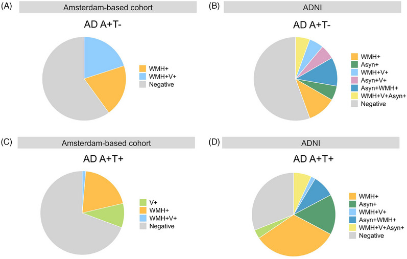 Figure 1
 Figure 2
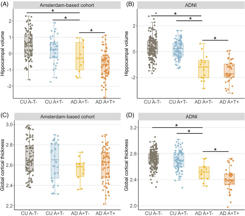 Figure 3
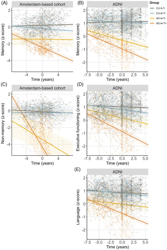 Figure 4
Peer Reviews
No public reviews on file for this paper yet. If you reviewed it on a platform where reviews are public (OpenReview, ICLR, NeurIPS, ICML), you can paste yours below so the community can read it here.
Videos
No videos yet. Explain this paper in a talk, walkthrough, or lecture? Add one.
Taxonomy
TopicsDementia and Cognitive Impairment Research · Neurological Disease Mechanisms and Treatments · Retinal Imaging and Analysis