Cone Beam Computed Tomography Evaluation of Stafne Bone Defect: A Case Series and Review of Radiographic Features
Ibrahim Yamany, Hanadi Sabban

TL;DR
This paper presents four cases of Stafne bone defects and shows how cone beam computed tomography helps in accurately diagnosing these rare, often asymptomatic lesions.
Contribution
The study highlights the diagnostic value of CBCT in identifying both typical and unusual presentations of Stafne bone defects.
Findings
CBCT provided detailed 3D imaging that helped distinguish SBDs from other mandibular pathologies.
Unusual features like anterior and bilateral SBDs were accurately identified using CBCT.
All cases confirmed SBDs as benign, supporting conservative management without treatment.
Abstract
Background: Stafne's bone defects (SBDs) are rare, intraosseous lesions not only localized in the mandible but also asymptomatic by default and found occasionally at radiographically investigations. The size and location of these defects can vary, although most are located in the posterior mandible. Since anterior variants are less frequently reported, diagnostic imaging is crucial for distinguishing SBDs from other diseases. This case series documents both familiar and unusual appearances, highlighting the diagnostic value of cone beam computed tomography (CBCT) in the evaluation of SBDs. Case Presentation: This study evaluated four instances of SBDs using CBCT. In Case 1, a 48-year-old man without any clinical symptoms had a characteristic posterior SBD located beneath the inferior alveolar canal. Case 2 described a 28-year-old woman's unusual anterior mandibular SBD, which was…
Genes, proteins, chemicals, diseases, species, mutations and cell lines named across the full text — each resolved to its canonical identifier and authoritative record.
Click any figure to enlarge with its caption.
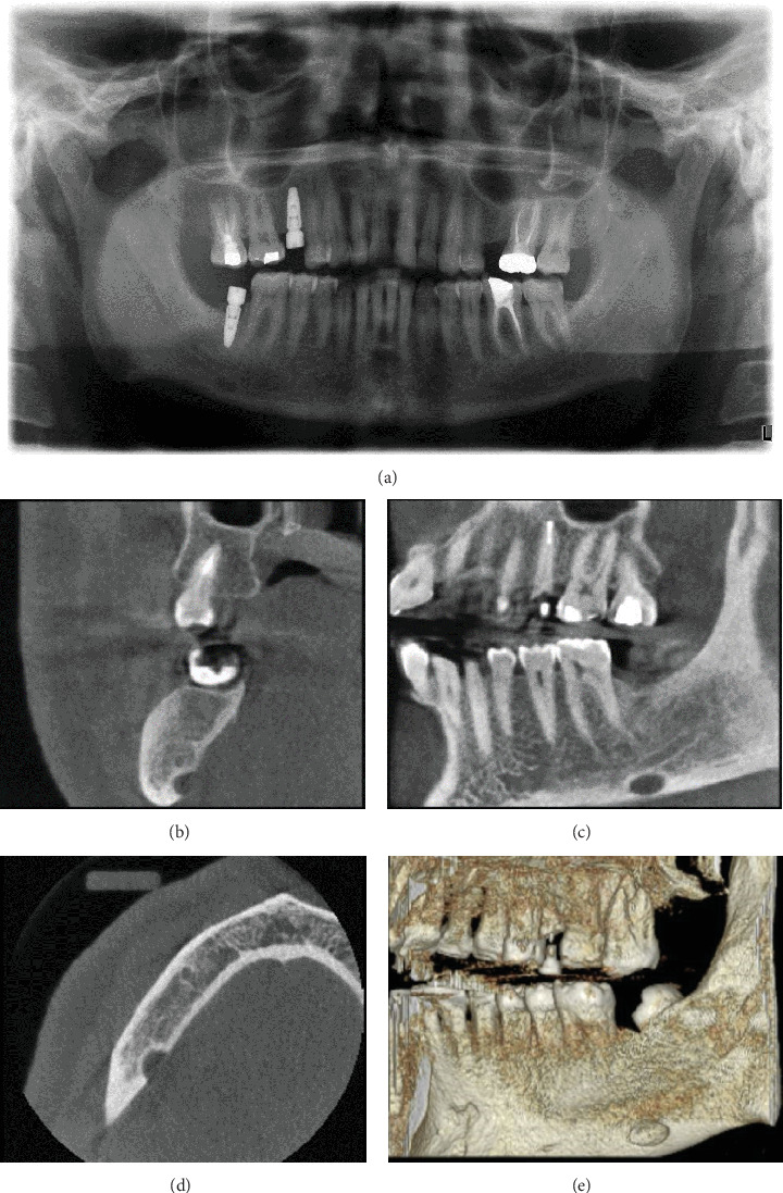 Figure 1
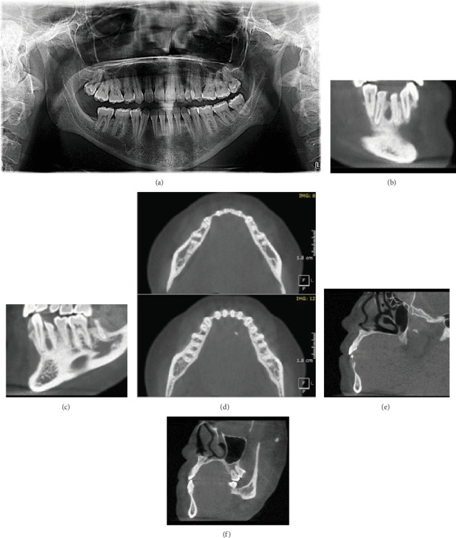 Figure 2
 Figure 3
 Figure 4
Peer Reviews
No public reviews on file for this paper yet. If you reviewed it on a platform where reviews are public (OpenReview, ICLR, NeurIPS, ICML), you can paste yours below so the community can read it here.
Videos
No videos yet. Explain this paper in a talk, walkthrough, or lecture? Add one.
Taxonomy
TopicsOral and Maxillofacial Pathology · Dental Radiography and Imaging · Bone Tumor Diagnosis and Treatments