Morphological Characterization of Intrafollicular Epithelial Bodies (IFEBs) in Rabbit Peyer’s Patches
Tiziana Tamborrino, Denise Bonente, Marì Regoli, Valentina Costa, Virginia Barone, Emiliana Giacomello, Giulia Collodel, Niccolò Fagni, Claudio Nicoletti, Eugenio Bertelli

TL;DR
This study describes newly identified structures called IFEBs in rabbit Peyer’s patches, showing they detach from the epithelium and migrate into lymphoid tissue.
Contribution
The paper introduces and characterizes intrafollicular epithelial bodies (IFEBs) as a novel feature of Peyer’s patch follicle-associated epithelium.
Findings
IFEBs display features of follicle-associated epithelium, including cytokeratin-positive cells and junction-associated molecules.
IFEBs detach from the epithelium and migrate into lymphoid tissue, losing their basement membrane and becoming non-polarized.
Serial sections confirm IFEBs form by separation from the FAE while maintaining epithelial integrity.
Abstract
Follicle-associated epithelium (FAE) covering the lymphoid follicles of Peyer’s patches (PPs) plays a central role in mucosal immunity. Here, we investigated FAE-derived intrafollicular epithelial bodies (IFEBs) that apparently detach from the FAE and sink deep into the lymphoid tissue of the PPs. Analysis of rabbit PP FAE was carried out by a variety of microscopy and immunohistochemistry techniques using a panel of specific antibodies to determine the nature of the IFEBs. IFEBs displayed the typical features of the FAE, with cytokeratin (CK)+ epithelial cells and CK+/vimentin+ M-cell-like cells. Serial sections of PP tissues showed that the IFEBs are formations frequently separated by the overlying FAE that maintains its integrity. Further, IFEBs showed the presence of junction-associated molecules like zonulin-1 and desmoplakins. Also, IFEBs apparently disaggregate within the…
Genes, proteins, chemicals, diseases, species, mutations and cell lines named across the full text — each resolved to its canonical identifier and authoritative record.
Click any figure to enlarge with its caption.
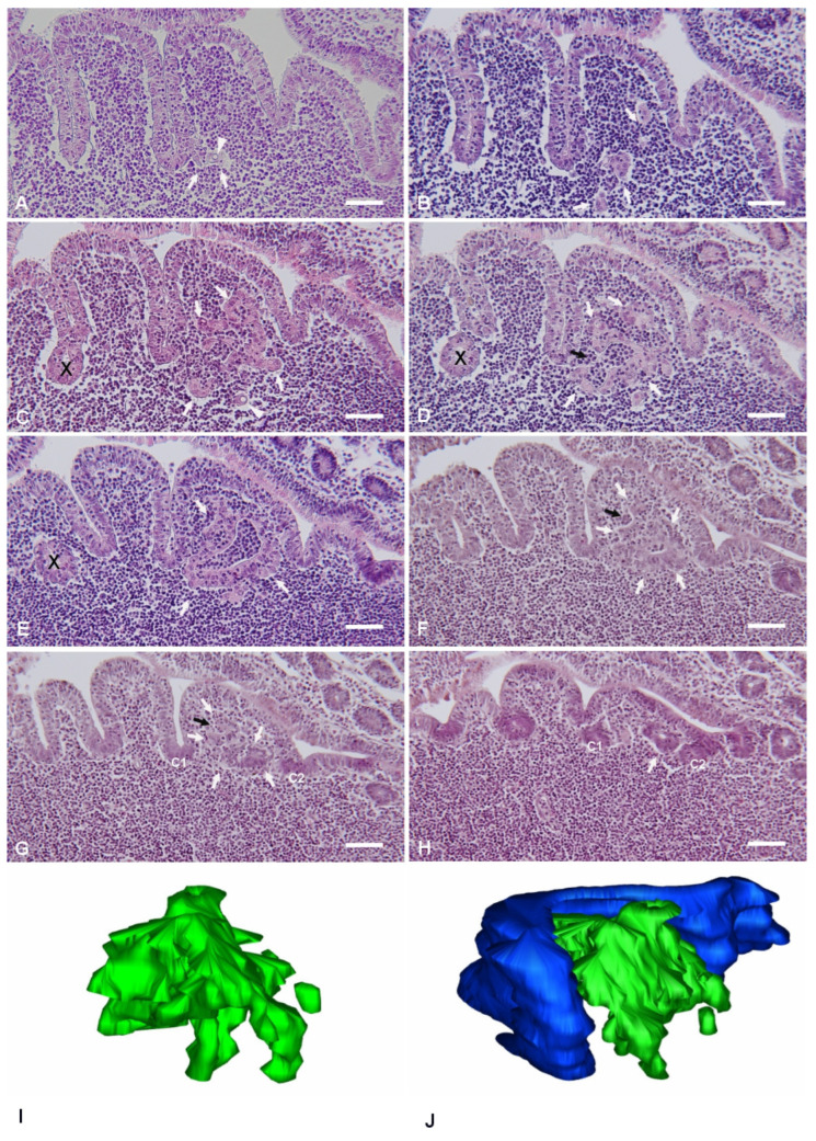 Figure 1
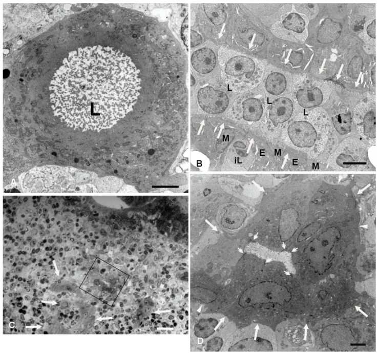 Figure 2
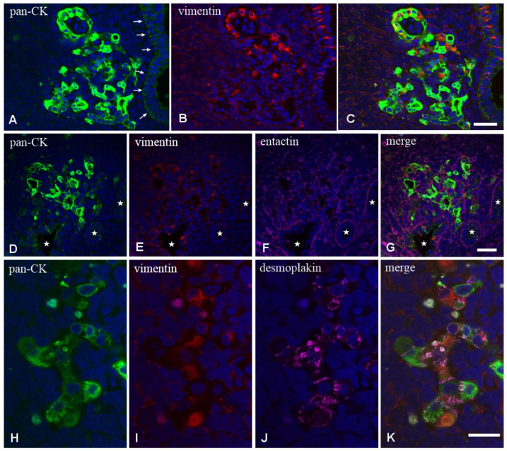 Figure 3
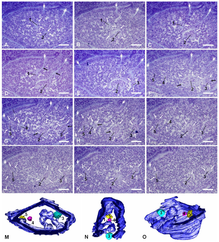 Figure 4
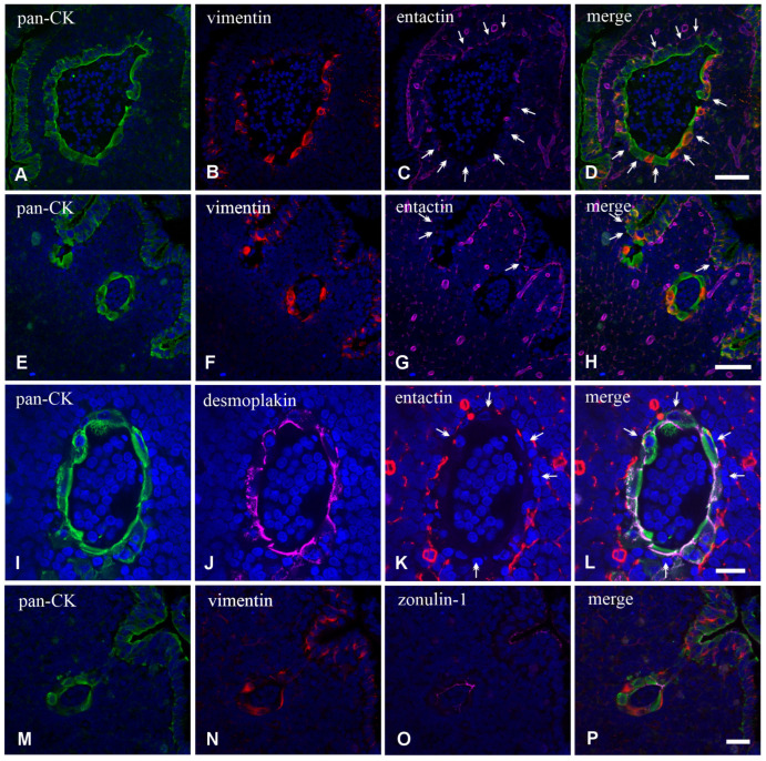 Figure 5
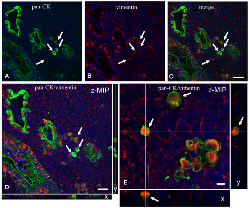 Figure 6
Peer Reviews
No public reviews on file for this paper yet. If you reviewed it on a platform where reviews are public (OpenReview, ICLR, NeurIPS, ICML), you can paste yours below so the community can read it here.
Videos
No videos yet. Explain this paper in a talk, walkthrough, or lecture? Add one.
Taxonomy
TopicsImmunotherapy and Immune Responses · Immune Response and Inflammation · Skin and Cellular Biology Research