Comparing Electromyographic Muscle Activities and Kinematics During Sit-to-Stand Transitions in Patients with Adult Spinal Deformity Versus Healthy Controls
Yukako Hayamizu, Tetsuyuki Nagafusa, Kumi Sasaki, Masaaki Nagashima, Katsuya Yamauchi, Tomohiko Hasegawa, Go Yoshida, Tomohiro Banno, Hideyuki Arima, Shin Oe, Tomohiro Yamada, Yukihiro Matsuyama, Yu Yamato

TL;DR
This study compares muscle activity and movement during sit-to-stand transitions in people with adult spinal deformity and healthy individuals, revealing differences in muscle use and motion.
Contribution
The study provides new insights into neuromuscular and biomechanical adaptations during sit-to-stand transitions in adult spinal deformity patients.
Findings
ASD patients showed increased muscle activation in the biceps femoris and soleus during sit-to-stand transitions.
ASD patients exhibited greater joint motion and longer phase durations compared to healthy controls.
The findings suggest neuromuscular and biomechanical differences in ASD patients during STS transitions.
Abstract
Background/Objectives: Adult spinal deformity (ASD) affects sit-to-stand (STS) transitions due to abnormal spinal alignment, influencing muscle function. This study investigated lower-extremity electromyographic activity and kinematic parameters during STS transitions in ASD patients. Methods: A cross-sectional study was conducted with ASD patients scheduled for corrective surgery. The STS task was divided into three phases, and electromyographic activity, temporal parameters, and joint kinematics were compared between ASD patients and controls. Surface electromyography measured muscle activity, and a high-speed camera recorded phase durations and joint movements. Results: Compared to 17 controls, 17 ASD patients exhibited significantly increased %MVIC (ASD, controls, p-value) in the biceps femoris during the flexion momentum phase (23.7 ± 26.5, 12.3 ± 8.6, p = 0.048) and extension…
Genes, proteins, chemicals, diseases, species, mutations and cell lines named across the full text — each resolved to its canonical identifier and authoritative record.
Click any figure to enlarge with its caption.
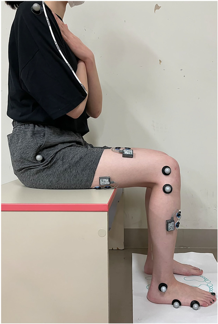 Figure 1
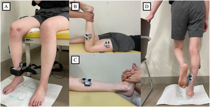 Figure 2
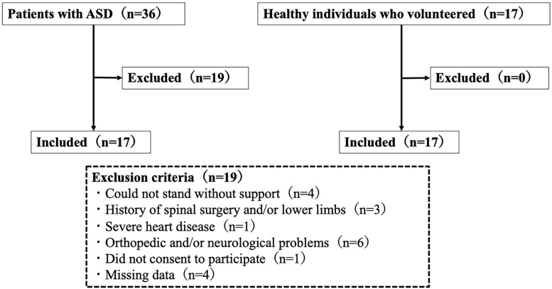 Figure 3
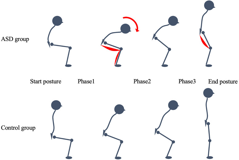 Figure 4
Peer Reviews
No public reviews on file for this paper yet. If you reviewed it on a platform where reviews are public (OpenReview, ICLR, NeurIPS, ICML), you can paste yours below so the community can read it here.
Videos
No videos yet. Explain this paper in a talk, walkthrough, or lecture? Add one.
Taxonomy
TopicsScoliosis diagnosis and treatment · Spinal Fractures and Fixation Techniques · Balance, Gait, and Falls Prevention