Complete genome sequence of Anaerococcus sp. strain AH8042_DFU013_CI05 isolated from a diabetes-related foot ulcer
Nan Hao, Sarah Vreugde, Robert Fitridge, Keith E. Shearwin

TL;DR
The complete genome of a new Anaerococcus strain from a diabetic foot ulcer is sequenced and described.
Contribution
The complete genome sequence of Anaerococcus sp. strain AH8042_DFU013_CI05 is reported for the first time.
Findings
The genome includes a 1,752,963 bp circular chromosome with 32% G+C content.
A 14,073 bp plasmid with 29% G+C content was also identified.
Abstract
This study presents the complete genome sequence of Anaerococcus sp. strain AH8042_DFU013_CI05, isolated from a diabetes-related foot ulcer at The Queen Elizabeth Hospital in Adelaide, Australia. The genome comprises a 1,752,963 bp circular chromosome and a 14,073 bp plasmid, with G+C contents of 32% and 29%, respectively.
Genes, proteins, chemicals, diseases, species, mutations and cell lines named across the full text — each resolved to its canonical identifier and authoritative record.
Click any figure to enlarge with its caption.
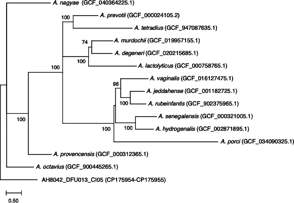 Fig 1
Fig 1| Parameters | Value |
|---|---|
| Number of contigs | 2 |
| Number of reads | 63,268 |
| N50 | 8,897 bp |
| Genome sizes: chromosome; plasmid | 1,752,963 bp; 14,073 bp |
| Coverage: chromosome; plasmid | 142 ×; 410 × |
| G + C content: chromosome; plasmid | 32%; 29% |
| Number of coding sequences (CDS) | 1,749 |
| Number of tRNA genes | 47 |
| Number of tmRNA genes | 1 |
| Number of rRNA genes | 12 |
| Number of ncRNA genes | 5 |
| Number of CRISPR loci | 1 |
| Accession numbers: chromosome; plasmid |
- —Aushealth
Peer Reviews
No public reviews on file for this paper yet. If you reviewed it on a platform where reviews are public (OpenReview, ICLR, NeurIPS, ICML), you can paste yours below so the community can read it here.
Videos
No videos yet. Explain this paper in a talk, walkthrough, or lecture? Add one.
Taxonomy
TopicsGenomics and Phylogenetic Studies · Plant Pathogens and Fungal Diseases · Oral microbiology and periodontitis research
ANNOUNCEMENT
The genus Anaerococcus, first proposed in 2001 with Anaerococcus prevotii as its type species (1), comprises 15 validly described species of gram-positive, strictly anaerobic bacteria listed in the LPSN (List of Prokaryotic Names with Standing in Nomenclature) (2). These species are frequently isolated from clinical specimens, including abscesses and infections at various body sites (3). Notably, Anaerococcus species are highly prevalent in diabetes-related foot ulcers (4–6), underscoring their potential role in the pathogenesis of these infections and highlighting the importance of sequencing to elucidate their contribution to wound microbiota.
A swab sample was collected from a diabetes-related ulcer on the left foot of a male patient at the Multi-disciplinary Foot Clinic, The Queen Elizabeth Hospital, Adelaide. Ethics approval and written informed consent were obtained prior to sample collection. After cleaning and debriding the wound, a swab was taken using the Levine technique (7), placed into Sigma Transwab Liquid Amies solution, and transferred to an anaerobic workstation. The sample was plated on tryptic soy agar supplemented with 5% defibrinated sheep blood and incubated anaerobically at 37°C for 3 days. Species identification using matrix-assisted laser desorption ionization-time of flight (MALDI-TOF) mass spectrometry revealed six known bacterial species and one unidentified species, designated AH8042_DFU013_CI05.
Strain AH8042_DFU013_CI05 was propagated anaerobically in the Brain Heart Infusion medium with 0.2% Tween 80 for 3 days at 37°C and harvested by centrifugation. Genomic DNA was extracted using the DNeasy Blood & Tissue Kit (Qiagen). A Nanopore sequencing library was prepared with the SQK-RBK114.96 Rapid Barcoding Kit and sequenced on a GridION sequencer using an R10.4.1 flow cell. Reads were base-called and demultiplexed with Guppy v4.2.0 in super-accurate (SUP) mode.
De novo assembly was performed with Flye v2.9.2-b1786 (8) following the Dragonflye v1.0.5 pipeline (Robert A. Petit III, Wyoming Public Health Laboratory). Assembly (Table 1) revealed a single circular chromosome (1,752,963 bp) and a plasmid (14,073 bp). The genome was annotated with Bakta v1.10.1 (db5.1.0) (9), identifying the features listed in Table 1. PHASTER (10) further predicted the presence of one intact prophage. Default parameters were used for assembly and annotation.
Ribosomal Multilocus Sequence Typing (rMLST) (11) identified AH8042_DFU013_CI05 as belonging to the genus Anaerococcus but failed to assign it to a specific species. Average nucleotide identity (ANI) analysis with FastANI v1.34 (12) revealed that the isolate shares 80.18% identity with the type strain Anaerococcus prevotii DSM 20548 (NCBI: ASM2410v1). A phylogenetic tree constructed with 14 Anaerococcus species from the LPSN indicated that AH8042_DFU013_CI05 is evolutionarily distinct from other known Anaerococcus species (Fig. 1).
Phylogenetic tree of Anaerococcus species constructed using kSNP4.1 (13) with an optimal k-mer size of 19 and 6,824 SNPs, employing a parsimony algorithm to minimize evolutionary steps. The tree was visualized in MEGA v11.0.13 (14), with genome accession numbers indicated in parentheses. Bootstrap values (100 replicates) are shown at nodes.
The reference list from the paper itself. Each links out to its DOI / PubMed record.
- 1Ezaki T, Kawamura Y, Li N, Li ZY, Zhao L, Shu S. 2001. Proposal of the genera Anaerococcus gen. nov., Peptoniphilus gen. nov. and Gallicola gen. nov. for members of the genus Peptostreptococcus. Int J Syst Evol Microbiol 51:1521–1528. doi:10.1099/00207713-51-4-152111491354 · doi ↗ · pubmed ↗
- 2Parte AC, Sardà Carbasse J, Meier-Kolthoff JP, Reimer LC, Göker M. 2020. List of prokaryotic names with standing in nomenclature (LPSN) moves to the DSMZ. Int J Syst Evol Microbiol 70:5607–5612. doi:10.1099/ijsem.0.00433232701423 PMC 7723251 · doi ↗ · pubmed ↗
- 3Murphy EC, Frick IM. 2013. Gram-positive anaerobic cocci--commensals and opportunistic pathogens. FEMS Microbiol Rev 37:520–553. doi:10.1111/1574-6976.1200523030831 · doi ↗ · pubmed ↗
- 4Dowd SE, Wolcott RD, Sun Y, Mc Keehan T, Smith E, Rhoads D. 2008. Polymicrobial nature of chronic diabetic foot ulcer biofilm infections determined using bacterial tag encoded FLX amplicon pyrosequencing (b TEFAP). P Lo S One 3:e 3326. doi:10.1371/journal.pone.000332618833331 PMC 2556099 · doi ↗ · pubmed ↗
- 5Smith K, Collier A, Townsend EM, O’Donnell LE, Bal AM, Butcher J, Mackay WG, Ramage G, Williams C. 2016. One step closer to understanding the role of bacteria in diabetic foot ulcers: characterising the microbiome of ulcers. BMC Microbiol 16:54. doi:10.1186/s 12866-016-0665-z 27005417 PMC 4804642 · doi ↗ · pubmed ↗
- 6Travis DJ, Bradbury J, Benkendorff K. 2023. Toward non-invasive collection methods for sampling the microbiome of diabetic foot ulcers. Int Wound J 20:3731–3737. doi:10.1111/iwj.1426737501084 PMC 10588311 · doi ↗ · pubmed ↗
- 7Angel DE, Lloyd P, Carville K, Santamaria N. 2011. The clinical efficacy of two semi-quantitative wound-swabbing techniques in identifying the causative organism(s) in infected cutaneous wounds. Int Wound J 8:176–185. doi:10.1111/j.1742-481X.2010.00765.x 21303456 PMC 7950681 · doi ↗ · pubmed ↗
- 8Kolmogorov M, Yuan J, Lin Y, Pevzner PA. 2019. Assembly of long, error-prone reads using repeat graphs. Nat Biotechnol 37:540–546. doi:10.1038/s 41587-019-0072-830936562 · doi ↗ · pubmed ↗