Aggregatibacter actinomycetemcomitans induces biofilm formation of Streptococcus sanguinis on titanium implants
Andrea Schubert, Jana Marisa Friebel, Oskar Bunz, Christoph Sasse, Ralf Bürgers, Torsten Wassmann

TL;DR
This study shows that Aggregatibacter actinomycetemcomitans boosts Streptococcus sanguinis biofilm formation on titanium implants, but surface roughness effects remain unclear.
Contribution
The study reveals a novel synergistic interaction between two bacterial species in biofilm formation on implant surfaces.
Findings
Dual-species biofilms of S. sanguinis and A. actinomycetemcomitans showed increased biofilm formation compared to single-species.
Surface roughness of titanium implants did not conclusively affect biofilm formation.
Fluorescence microscopy confirmed the efficiency of washing steps in biofilm analysis.
Abstract
This study aims to investigate the distinct behaviors of single-species and dual-species biofilms formed by Streptococcus sanguinis and Aggregatibacter actinomycetemcomitans on different titanium and implant surfaces. Four types of surfaces were examined: two clinically used implant surfaces, a super-polished surface and a sand-blasted surface of grade 4 titanium. Specimens were incubated with single- and dual-species biofilms for 24 h. Biofilm formation was determined based on the amount of total DNA extracted from the bacteria. In order to specifically determine the biofilm formation of Streptococcus sanguinis, qPCR experiments were carried out. Staining followed by fluorescence microscopy was employed to verify the efficiency of the washing steps. Biofilm formation by single- and dual-species cultures was observed on all tested implant surfaces. However, a clear influence of…
Genes, proteins, chemicals, diseases, species, mutations and cell lines named across the full text — each resolved to its canonical identifier and authoritative record.
Click any figure to enlarge with its caption.
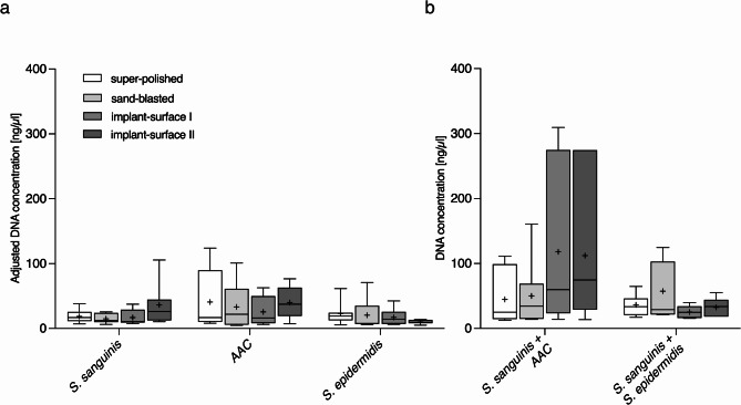 Figure 1
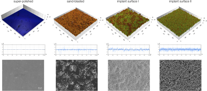 Figure 2
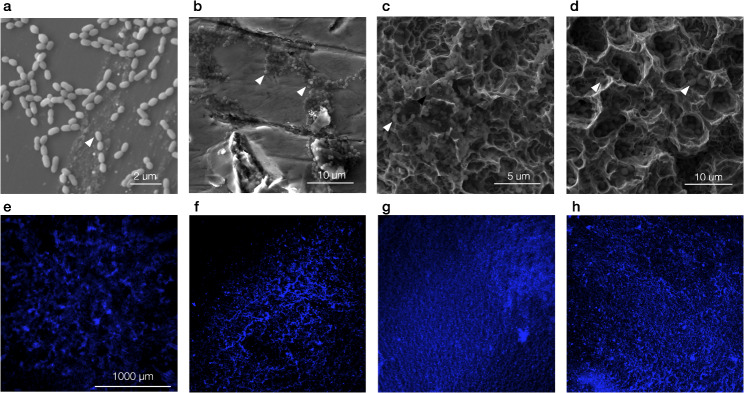 Figure 3
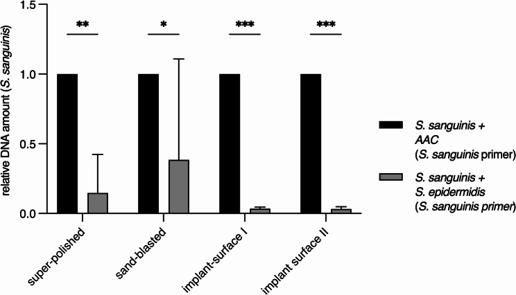 Figure 4
- —Herzzentrum Göttingen (1018)
Peer Reviews
No public reviews on file for this paper yet. If you reviewed it on a platform where reviews are public (OpenReview, ICLR, NeurIPS, ICML), you can paste yours below so the community can read it here.
Videos
No videos yet. Explain this paper in a talk, walkthrough, or lecture? Add one.
Taxonomy
TopicsDental Implant Techniques and Outcomes · Dental materials and restorations · Oral microbiology and periodontitis research
Background
Peri-implantitis describes the inflammation of the tissues adjacent to osseointegrated implants that results in the loss of peri-implant bone tissue [1, 2]. Peri-implantitis shows high prevalence and is considered a major cause of implant loss [2, 3]. Microbiologically, it represents a biofilm-associated disease with a variable microbial spectrum [4, 5]. For preventive considerations the minimization of microbial adhesion on implant materials is desirable. In addition to the composition of the implant material, microbial adhesion depends on surface charge, surface free energy (SFE) and surface roughness [6–8].
Ti-6Al-4 V is a titanium alloy commonly used in dental implantology as an implant material per se and as a material for prosthetic abutments with evidence-based long-term reliability [9]. Sand-blasted and acid-etched surface modifications are used to optimize osseointegration, resulting in average mean roughness values (R_a_) of 1 to 2 μm [10]. The adhesion of biofilms to titanium surfaces with different roughness values has been investigated in vitro and in vivo: above an R_a_ threshold of 0.2 μm adhesion correlates with increasing roughness [11, 12]. In the pathogenesis of peri-implantitis, gram-positive early colonizers of oral surfaces such as S. sanguinis are considered non-pathogenic, whereas gram-negative species such as AAC and Eikenella corrodens seem to be associated with peri-implant inflammation [13–15]. The adhesion behavior of different microorganisms in monospecies biofilms on dental materials differs: the adhesion of Candida albicans showed a stronger positive correlation with increasing roughness values than S. sanguinis [16]. Little is known about how the adhesion behavior of different microorganisms in a multi-species biofilm changes depending on surface roughness. A changed biofilm composition may influence the pathogenicity with regard to the development of peri-implantitis. Although there is evidence that implant surfaces with low roughness do not prevent peri-implantitis [17], investigations on the influence of implant surfaces on peri-implant diseases are scarce [18].
The present in vitro study investigated the proportionate composition of dual-species biofilms from a non-pathogenic (S. sanguinis) and AAC, a microorganism that is often associated with peri-implantitis [19, 20], on titanium surfaces with different surface treatments and correspondingly different surface roughness. The results of the present study make a basic scientific contribution to the knowledge about biofilm formation on dental implant surfaces.
Materials and methods
Specimen preparation
Cylindrical specimens (10 mm x 2 mm) of four titanium materials with different surface specifications were included in the present study: super-polished specimens of grade 4 titanium (‘super-polished’), sand-blasted specimens of grade 4 titanium (‘sand-blasted’) and specimens of two commercially available and clinically used titanium implant surfaces (‘implant surface I’: Myplant Two titanium implants, Hager & Meisinger GmbH, Neuss, Germany; ‘implant surface II’: Semados titanium implants, BEGO GmbH & Co. KG, Bremen, Germany).
Super-polished specimens were manufactured as follows: rods of grade 4 titanium (Hempel metals and more, Duebendorf-Zurich, Switzerland) were sliced into disks using a separating machine (Micracut 201, Metkon, Bursa, Turkey) and polished with an automated grinding machine (Digiprep 251, Metkon) and silicon carbide grinding paper with decreasing grain sizes (400, 800, 1200, 4000), then polished using polishing papers with grit sizes of 3 μm and 1 μm. Sand-blasted specimens were obtained likewise, but sand-blasted instead of polished using an aluminum oxide grit size of 110 μm (Alublast 110 μm, Henry Schein Inc., Melville, USA) for 6 s at a pressure of 1 bar and at a distance of 100 mm using a sandblasting device (Basic Master, Renfert GmbH, Hilzingen, Germany). Specimens of implant surface 1 and implant surface II were provided by the respective implant manufacturers.
Surface characterization
Mean surface roughness R_a_ was calculated for five specimens of each material via confocal microscopy (Zeiss Smartproof 5, Carl Zeiss Microscopy, Jena, Germany) and automated software analysis (Confomap, Carl Zeiss Microscopy). The SFE was determined as described before [16]. In brief, contact angle measurements were performed for each tested material: one µl of distilled water and methylene iodide, respectively, was applied to the specimen’s surface. Within 30 s after application, a computer-aided measurement device (Drop Shape Analyzer DSA25, Krüss, Hamburg, Germany) performed ten contact angle measurements for each liquid. The SFE was calculated using the formula introduced by Owens and Wendt [21].
Scanning electron microscopy
The imaging of the test specimen surfaces was performed using a scanning electron microscope (JEOL JSM-IT500 InTouchScope, JEOL Ltd., Akishima, Japan) at a voltage of 20 kV. For each test material, one representative area with and without bacterial colonization was captured at a magnification of 2700x.
Microbial adhesion
S. sanguinis (lot nr. 20068, Deutsche Sammlung von Mikroorganismen und Zellkulturen DSMZ, Braunschweig, Germany), AAC (lot nr. 8324, DSMZ) and Staphylococcus epidermidis (lot nr. 8340, DSMZ) were cultured in liquid Brain Heart Infusion Broth medium (lot nr. CM1135, Oxoid Limited, Hampshire, UK) at 37 °C. Microorganisms were harvested by centrifugation, washed twice with phosphate-buffered saline (PBS, Merck, Darmstadt, Germany) and resuspended in PBS. For experimental procedures, suspensions in PBS with an adjusted optical density of 0.2 at 600 nm were prepared by densitometry (Bio Photometer, Eppendorf, Hamburg, Germany). Under sterile conditions, specimens of the test materials were transferred to 24-well plates and attached to well bottoms using silicone (Z-Dupe, Henry Schein Dental, Langen, Germany). For the mono-species biofilms, 1 ml of S. sanguinis, S. epidermidis or AAC suspension was added to each well and incubated for 24 h at 37 °C and 55 rpm.
In order to obtain a similar bacterial density as in the single culture, the amount was halved in the two-species biofilm and later adjusted mathematically in the analysis. For the dual-species biofilms, 500 µl of each bacterial suspension was added to the well, mixed using a sterile pipette (Sarstedt AG & Co. KG, Nürnbrecht, Germany) and incubated under the previously described conditions.
Fluorescence staining
In order to visualize the bacterial biofilms on the test surfaces, fluorescence staining was performed using the DNA dye Hoechst 33,342 (Sigma-Aldrich, St. Louis, USA). Following the incubation, specimens were washed three times with 0.85% saline; 1 mL bisbenzimide H 33,342 trihydrochloride (Sigma Aldrich, Munich, Germany) was added to each specimen for 10 min. Staining solution was removed via three washing steps with 0.85% saline, and microbial cultures were fixated using 8% paraformaldehyde solution. After 10 min, specimens were dried and mounted on object slides for visualization via fluorescence microscopy (BZ-X710, Keyence, Osaka, Japan).
Biofilm removal and DNA isolation
The biofilm removal and isolation of bacterial DNA was performed using the QIAamp DNA Mini Kit (Qiagen N.V., Venlo, Netherlands). After incubation, the bacterial suspension was removed, specimens were washed with 1 ml 0.85% saline and transferred to sealable reaction tubes (Eppendorf, Hamburg, Germany). The cell lysis and isolation of the DNA was conducted in accordance with the specifications provided by the manufacturer. The removal of biofilms was confirmed by repeated fluorescence staining following the described protocol.
Primer designing and real-time PCR
Specific oligonucleotide primers were identified for use in real-time PCR with the selected bacterial strains based on literature research in the National Center for Biotechnology Information (NCBI) database. The sensitivity and specificity of the identified primer sequences were assessed using the Basic Local Alignment Search Tool (BLAST, http://blast.ncbi.nlm.nih.gov/Blast.cgi) in an in-silico PCR. Primer sequences lacking specificity for a certain bacterial strain or cross-reactivity with two bacterial strains were excluded. After a data-based review of the sequences, a Genomics product manufacturer (Eurofins Genomics, Ebersberg, Germany) produced an individual primer for each bacterial species (Table 1).
qPCR experiments were performed in a Mic qPCR Cycler (Bio Molecular Systems, Upper Coomera, Australien) according to the standard protocol. Briefly, a denaturation step at 94 °C after an initialization step at 95 °C was used. The denaturation temperature was 57.5 °C, based on the primer sequence. A total of 30 cycles were used for the analysis. Primers specific for S. sanguinis were used for all qPCR experiments (Table 1). A SYBR Green mix (Kapa SYBR FAST, Roche Holding AG, Basel, Schweiz) was used according to the user’s manual. Data analysis was performed using the Mic qPCR Analysis Software (Bio Molecular Systems, Upper Coomera, Australien). Five biological replicates were performed. After the real-time PCR, an exemplary reaction product for each primer was sterilely stored at -18 °C and sent to an external analysis laboratory for DNA sequencing.
Table 1. Overview of oligonucleotide primers (target species and sequence of the species-specific forward (F) and reverse primer (R).)Target speciesprimerprimer sequence S. sanguinis S2F: CAA AAT TGT TGC AAA TCC AAA GGR: GCT ATC GCT CCC TGT CTT TGA
Statistical analyses
The adhesion experiment data, including DNA concentrations, are presented as boxplots with 75% quartile, median and 25% quartile. To ensure a comparable representation of DNA concentration, the values for the single-species biofilms were adjusted accordingly. During the experiment, the bacterial strains in the two-species biofilms were halved to achieve a bacterial density comparable to that of the single-species biofilms. To account for these differences in bacterial quantities, the results were also halved for Fig. 1a and labeled as “adjusted values.”
Fig. 1DNA concentration of the investigated biofilms on varying test materials. (a) Adjusted (halved) single-species biofilms and (b) measured DNA concentrations of two-species biofilms. The + marks mean values, 75% quartiles, median and 25% quartiles are shown
The qPCR evaluation comparing dual-species biofilms of S. sanguinis with AAC or with S. epidermidis were assessed for normal distribution using a Q-Q plot and then analyzed using a two-way ANOVA with Šídák’s multiple comparisons test, with an alpha level of 0.05.
Results
Surface characteristics
The R_a_ values of the examined specimens ranged from 0.062 μm (± 0.023, super-polished) to 1.405 μm (± 0.093, implant surface I). SFE values varied from 55.430 mN/m (± 2.861, super-polished) to 65.214 mN/m (± 4.577, implant surface I) (Table 2). No correlation was found between surface roughness and SFE (data not shown).
Figure 2 provides an overview of the 3D representation of the surface topography, the 2D representation of the roughness profiles and the corresponding scanning electron microscopy images. Super-polished specimens had a macroscopically smooth appearance corresponding to a low R_a_ value, while the higher peaks in the profiles of the other materials reflected higher R_a_ values. The surface of the sand-blasted group showed occasional very high peaks in both the three-dimensional representation and the two-dimensional profile curve.
Fig. 2. Surface characteristics of the tested materials. 3D representation of the surface topography (upper row), 2D representation of the roughness profile (middle row), scanning electron microscopy (bottom row). The super-polished surface appears homogenous, the sand-blasted surface and implant surfaces I and II are irregular
The scanning electron microscopy images unveiled that the super-polished sample group had a homogeneous surface without grooves or ridges (Fig. 2). The sand-blasted sample group appeared rugged and rough due to the impacts of the blasting material, with the structuring of the sand-blasted surface appearing significantly coarser compared to implant surfaces I and II. In addition to coarse inhomogeneities, the surfaces of implant surfaces I and II exhibited a finely rugged surface structure, possibly due to the two-step surface treatment involving sandblasting and acid etching. Compared to implant surface II, implant surface I had a similar but finer surface structure.
Table 2. Mean values and standard deviations of R_a_ and SFE values of the tested materialsTest materialRa [µm]Surface free energy [mN/m]super-polished0.062 (± 0.023)55.43 (± 2.86)sand-blasted0.505 (± 0.232)63.93 (± 1.96)implant surface I1.325 (± 0.138)65.21 (± 4.58)implant surface II1.405 (± 0.093)59.01 (± 2.93)
Biofilm formation
In order to find out whether the surface roughness of titanium implants has an influence on biofilm formation with various bacteria, corresponding investigations were carried out. Therby two typical oral bacteria species (AAC and S. sanguinis) and one non typical oral bacterial species (S. epidermidis) were used. Fluorescence staining and scanning electron microscopy were performed to visualize the adhesion of mono-species and dual-species to the tested materials. Exemplary images are shown in Fig. 3. The cells of S. sanguinis were organized in typical chains, while the cells of AAC were arranged in pairs or clusters. The cells of S. epidermidis formed clusters on the surface.
Fig. 3. Biofilm formation on the tested materials. Scanning electron microscopy (a-d) and fluorescence staining (e-h) of investigated biofilms on different test materials S. sanguinis on the super-polished surface (a), S. epidermidis on the sand-blasted surface I (b), S. sanguinis with AAC on implant surface I (c), S. sanguinis with S. epidermidis on implant surface II (d), AAC on the super-polished surface (e), S. sanguinis on implant surface I (f), S. epidermidis on implant surface II (g), S. sanguinis with AAC on implant surface II (h)
The concentration of isolated DNA was used for determination of the biofilm formation. Different colonization behaviour was observed in the tested bacteria, with the strongest biofilm formation being measured for AAC. However, no clear differences were found between the used titanium surfaces with regard to colonization of the various individual bacterial cultures (Fig. 1a). In contrast, the combination of S. sanguinis and AAC showed a higher amount of DNA on impant-surface I and II compared to the single cultures, whereas the co-cultivation of S. sanguinis and S. epidermidis did not lead to this strong tendency (Fig. 1a and b). DNA quantity of the mixed culture of S. sanguinis and S. epidermidis appears to correspond somewhat to the sum of the quantities of the corresponding individual cultures. Nevertheless, no significant differences could be observed. Based on these results the surface roughness of titanium implants appears to have no effect on biofilm formation under the tested conditions. In contrast, the composition of the bacterial cultures seems to influence directly the biofilm formation (Fig. 1b).
Quantification of S. sanguinis in two-species biofilms
We wanted to find out whether the biofilm formation of S. sanguinis can also be increased depending on certain microorganisms. Previous work has shown that co-cultivation of S. mutans and AAC increases biofilm formation [22], whereas a similar effect has not been described for S. epidermidis. By using AAC and S. epidermidis, it should be found out whether an increased biofilm formation of S. sanguinis can be induced in dependency of AAC. S. epidermidis was used as control. Therefore, the relative amount of S. sanguinis within dual-species biofilms, consisting of AAC or S. epidermidis, was investigated by qPCR (Fig. 4). Therefore, S. sanguinis specific primers were used (see method part). Five biological replicates were performed and data were statistical anaylsed using the two way ANOVA.
The two-species biofilm consisting of S. sanguinis and AAC shows that S. sanguinis was significantly more present in comparison to the mixed culture of S. sanguinis and S. epidermidis. This effect could be oberserved for all tested material surfaces (Fig. 4).
These data implies that AAC but not S. epidermidis supports biofilm formation of S. sanguinis on surfaces of titanium dental implants independent of the R_a_ value of the surface.
Fig. 4AAC increases the biofilm formation of S. sanguinis on the tested materials. Relative DNA amount of S. sanguinis for two-species biofilms on tested titanium surfaces, using S. sanguinis-specific primers. Samples with AAC and S. sanguinis were normalized to 1. Five biological replicates were used for analysis. Error bars indicated the standard deviation (SD). Significant differences between biofilm groups are indicated by asterisks (* = p ≤ 0.05; ** = p ≤ 0.001; **0 = p ≤ 0.0001)
Discussion
A critical point in the pathogenesis and etiology of peri-implant disease is the initial adhesion of bacteria [4, 5]. While the microbiome on an implant surface with healthy peri-implant tissue resembles a biofilm on the natural tooth substance, peri-implant lesions are dominated by gram-negative, parodontopathogenic pathogens such as AAC, P. gingivalis or T. denticola [13–15, 22]. Thus, the species-specific composition of peri-implant biofilms is of crucial importance in the etiology of peri-implant diseases.
Peri-implantitis normally occurs under anaerobic conditions. In this study, the biofilm experiments were carried out under aerobic conditions. This was done to obtain a simple, robust set up and to minimize deviations that are more likely to occur under anaerobic conditions. Of course, this can have an influence on biofilm formation. However, our results show that AAC increases biofilm formation in S. sanguinis, which is consistent with the data from Szafrański et al. [23]., where the experiments were performed in presence of CO_2_.
The mechanisms leading to the specific dominance of certain potentially pathogenic species are not yet understood. In this study we investigated titanium implants, which are still considered the gold standard in dental implantology [10, 24, 25], with regard on biofilm formation based on roughness and bacterial species.
The R_a_ value has already been discussed in various studies as a significant influence factor on bacterial adhesion [12, 26, 27], whereby surfaces with a higher R_a_ value have an increased potential for bacterial adhesion [12, 28]. In this work, a tendency was observed that biofilm formation of single-species cultures is independent of the surface roughness. This would partially explain why biofilms usually consist of several different microorganisms to increase the biofilm formation on different surfaces. This hypothesis is supported by our observations where we were able to recognize a dependency on the surface quality based on the measured DNA quantity for the mixed cultures. The biofilm formation on the implant-surface I and II, indicated by a higher amount of total bacterial DNA, seems to be increased in the mixed cultures of AAC and S. sanguinis than in the single cultures. This effect was not observed in the co-cultures of S. sanguinis and S. epidermidis. Therefore, biofilm formation seems to be more dependent on the bacterial composition than on the implant surface (Fig. 1b). Thus, cross-talk between different microorganisms from the same environment enables efficient colonization. However, this effect could not be confirmed by qPCR analysis of S. sanguinis. The data showed no significant increase in the amount of S. sanguinis in the mixed cultures with the different implants. Based on the current results, it is therefore not possible to make a definitive statement regarding biofilm formation on titanium implants in dependency on the R_a_ value. In future, further experiments are required in which bacteria from the oral microbiome are incubated in various combinations with titanium implants in order to gain a better understanding of oral biofilm formation in correlation with the R_a_ value.
The combination of S. sanguinis and AAC increases the biofilm formation in comparison to the corresponding single-species cultures. A similar effect was already described for AAC and S. mutans [23]. They could show that AAC activates quorum sensing (QS) in S. mutans by inducing the expression of the alternative sigma-factor encoding gene sigX which results in increased biofilm formation [23, 29, 30]. Although the regulatory mechanism for QS seems to be conserved in Streptococci [29], the knowledge about the crosstalk between S. sanguinis and AAC, leading to an enhanced biofilm formation, is still limited. Therefore, future works have to be done to shed light in the regulation of biofilm formation of AAC and S. sanguinis and typical microorganism involved in peri-implantitis [31].
Our results revealed that AAC increases the amount of S. sanguinis during biofilm formation on titanium surfaces. However, only S. sanguinis was quantified via qPCR, which means that no statement can be made about the amount of AAC within the biofilm. Thus, we cannot exclude whether the amount of AAC is changed under the tested conditions as well. Szafranski et al. already showed, that the cell number of AAC is not significantly increased in presence of S. mutans [23], but Sliepen et al.. demonstrated that the colonization of AAC on epethelium cells in presence of S. sanguinis can be inhibited [32]. We have studied biofilm formation on implants and not in connection with cell tissues. Therefore, we assume that only the biofilm of S. sanguinis, indicated by the increased amount of DNA, and not of AAC is affected during biofilm formation.
Conclusions
We demonstrate that beside the known inducing effect of AAC on biofilm formation of S. mutans [23], this microorganism also leads to increased biofilm formation of S. sanguinis by a so far unknown mechanism. This shows that AAC has a broad spectrum for cell-cell communication within the oral microbiome and is therefore a decisive factor for biofilm formation on implant surfaces within the oral cavity.
Although AAC is discussed controversial as a typical representative of peri-implantitis, our experiments have shown that this microorganism can induce Streptococci in general, regardless of whether they are early or late colonizers, and that it can therefore be induced in the early stages of peri-implantitis. Therefore, the early biofilm formation in correlation with peri-implantitis seems to require the cross-talk of microorganism for a successful coloization.
Future experiments need to be performed to provide a deeper insight into the crosstalk of microorganisms within the oral microbiome and their influence on biofilm formation of surface structures.
The reference list from the paper itself. Each links out to its DOI / PubMed record.