Clinical Presentation and Surgical Management of a Grynfelt Hernia: Report of a Clinical Case and Literature Review
Pabel Ruben Carbajal Cabrera, Ruben Daniel Pérez López, Yunuen Ailyn Morales Tercero, Itzel Ocampo Barrero

TL;DR
This paper reports a rare case of Grynfelt's hernia in a young woman and describes its successful surgical treatment.
Contribution
The paper presents a clinical case of Grynfelt's hernia with no prior surgical or traumatic history and discusses its management.
Findings
The patient underwent successful elective left lumbotomy surgery with hernioplasty using a polypropylene mesh.
No recurrence or complications were observed during follow-up at 20 days, 1, 3, and 6 months.
Surgical management decisions were based on intraoperative findings due to the lack of specific guidelines for this rare condition.
Abstract
Background: Grynfelt's lumbar hernia is the rarest of all abdominal wall hernias, accounting for between 1.5% and 2% of cases, with only 300–350 instances described to date. Lumbar hernias can be congenital or acquired, often triggered by trauma or surgery (iatrogenic). Diagnosis is clinical and confirmed via computed tomography. Surgical intervention is required for resolution, with repair performed either through open or laparoscopic surgery. Material and Methods: We present the case of a young female with no prior surgical or traumatic history, in whom the diagnosis of Grynfelt's hernia was made. Results: The patient underwent elective left lumbotomy surgery with hernioplasty using a supra-aponeurotic polypropylene mesh. Postsurgical recovery was adequate, and she was discharged 4 h after surgery. Follow-up in the general surgery outpatient clinic occurred at 20 days, 1, 3, and 6…
Genes, proteins, chemicals, diseases, species, mutations and cell lines named across the full text — each resolved to its canonical identifier and authoritative record.
Click any figure to enlarge with its caption.
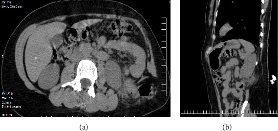 Figure 1
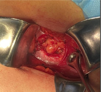 Figure 2
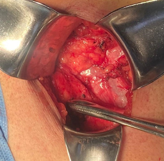 Figure 3
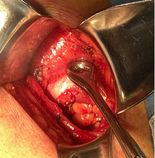 Figure 4
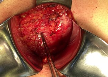 Figure 5
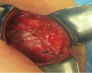 Figure 6
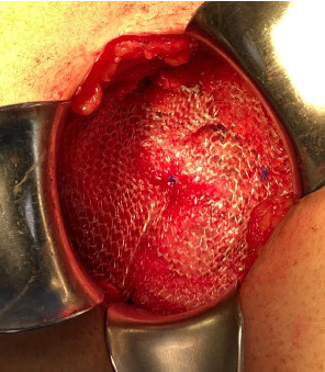 Figure 7
- —Universidad Nacional Autónoma de México
Peer Reviews
No public reviews on file for this paper yet. If you reviewed it on a platform where reviews are public (OpenReview, ICLR, NeurIPS, ICML), you can paste yours below so the community can read it here.
Videos
No videos yet. Explain this paper in a talk, walkthrough, or lecture? Add one.
Taxonomy
TopicsHernia repair and management · Congenital Diaphragmatic Hernia Studies · Gastrointestinal disorders and treatments
1. Introduction
A lumbar hernia is the protrusion of intraperitoneal or extraperitoneal contents through a defect in the posterolateral abdominal wall. These hernias are classified based on the anatomical triangle affected, first described in 1783 and 1866, respectively, and are named after the Jean–Louis Petit triangle (lower) [1] and the Grynfeltt–Lesshaft triangle (upper) [2].
With ~300–350 cases published in the literature, lumbar hernias represent 1.5%–2% of all abdominal hernias, with Grynfelt's hernia being more frequent [3]. These hernias are classified as either congenital or acquired. Approximately 20% are congenital, mainly due to defects in embryonic development, while 80% are acquired [4].
The Grynfeltt–Lesshaft triangle is an inverted triangle with boundaries formed superiorly by the twelfth thoracic rib, medially by the erector spinae and quadratus lumborum muscle group, and laterally by the internal oblique muscle [5]. The floor of this triangle is formed by the aponeurosis of the transversus muscle, and the roof is formed by the Latissimus Dorsi muscle and external oblique muscle [5].
Grynfeltt's triangle has three described weakened areas: immediately below the rib, where the transversalis fascia is not covered by the external oblique muscle; in the area where the twelfth dorsal intercostal neurovascular pedicle penetrates the fascia; and between the lower edge of the rib and Henle's ligament [6].
The aim of this article is to present a case report of a female patient with a congenital Grynfelt hernia, which has a very low prevalence. We describe the diagnostic approach and surgical treatment of Grynfelt's hernia.
2. Case Presentation
A 36-year-old female patient with no personal medical history. She denies smoking and has no history of other surgeries or trauma related to the lumbar region.
She presented to the Emergency Room reporting the onset of her current condition 2 years ago, characterized by intermittent stabbing pain located in the left lumbar region, associated with a left lumbar swelling that increased in size with the Valsalva maneuver. No other significant clinical symptoms were noted. On physical examination, a swelling was observed in the left posterolateral lumbar region, below the twelfth rib, measuring ~7 × 5 cm. The swelling showed no local color changes, increased in size with the Valsalva maneuver, was reducible, and soft to palpation. It was classified as a type A hernia according to the classification by Moreno-Egea et al. [7, 8].
After diagnosing and classifying the condition as a Grynfelt hernia, the patient was initially treated with NSAIDs. However, there was no clinical improvement, and further interventions were considered. A contrast-enhanced abdominal computed tomography (CT) scan revealed an aponeurotic defect located in the left lumbar region at the Grynfeltt–Lesshaft tract, measuring 69 × 51 mm, with a hernial sac containing retroperitoneal fat (Figure 1a,b).
Elective surgery was scheduled. The procedure was performed under regional anesthesia. The patient was placed in the right lateral decubitus position, and asepsis of the abdominal and lumbar regions was performed using 2% chlorhexidine. A lumbotomy was performed 6 cm below the twelfth rib, with an incision ~8 cm long. The latissimus dorsi and quadratus lumborum muscles were identified, and the external oblique muscle was dissected from the internal oblique muscle (Figures 2?–4). A 3 × 3 cm aponeurotic defect was found in the transverse abdominal muscle, along with a hernial sac containing retroperitoneal fat (Figure 5) [8]. Five centimeters of “healthy” aponeurosis were dissected around the periphery. The hernial sac was reduced without complications.
Plication of the internal oblique muscle onto the transversus abdominis muscle was performed horizontally using 1-0 polypropylene suture in simple interrupted stitches (Figure 6). A light polypropylene mesh, measuring 8 × 8 cm, was applied and fixed supraponeurotically to the external oblique with 2-0 polypropylene suture (Figure 7). The skin was closed with 3-0 nylon, and no surgical drains were placed.
The surgery was completed in 60 min without incidents or complications. The patient had an uneventful postoperative course and was discharged 4 h after the surgery.
Follow-up was conducted in the general surgery outpatient clinic at 20 days, 1, 3, and 6 months postoperatively, with no recurrence, complications, or other incidents.
3. Discussion
Grynfeltt's hernias are relatively rare, occurring in the lumbar region through a defect in the superior lumbar triangle, also known as the Grynfeltt–Lesshaft triangle. According to Hafner, Wylie, and Brush [9], a general surgeon will likely only have the opportunity to repair one case of lumbar hernia in their lifetime.
Barbette first proposed the presence of these hernias in 1672, while the initial documented mention was made by de Garangeot [10] in 1731. Ravaton [11] in 1750 performed the first surgical intervention for a strangulated lumbar hernia in a pregnant woman. Petit [1] in 1783 defined the anatomical boundaries of the inferior lumbar space, whereas Grynfeltt [2] in 1866 described the superior space. In 1890, Macready documented 25 cases, including two in the superior lumbar space, coining it as the “triangle of Grynfeltt–Lesshaft.” In 1916, Goodman highlighted the prevalence of hernias in the inferior space; however, studies after 1920 indicated a higher occurrence in the superior location [8, 12]. The first laparoscopic lumbar hernia repair was reported by Burick et al. [13, 14].
Congenital presentation is rare (20%), while acquired lumbar hernias (80%) are more frequent, often resulting from incisional or traumatic origins [7]. Of the acquired defects, 55% are spontaneous or primary, and the remaining 25% are secondary [13]. Several risk factors have been described, including age (generally between 50 and 70 years), obesity, extreme thinness, cachexia, chronic wasting disease, muscle atrophy, chronic bronchitis, infected wounds, and postoperative sepsis [6]. Old age and weight loss may also contribute [15].
The diagnosis of lumbar hernia is usually based on clinical suspicion, depending on physical examination findings [16]. These hernias are often asymptomatic, though patients may sometimes report back pain, especially in the flank or lower back, and a sensation of increased bulk [7]. The size of the hernia typically increases progressively, depending on the contents, which may include retroperitoneal fat, kidney, colon, omentum, spleen, or, in very rare cases, small intestine [7]. In cases of strangulation, nausea, vomiting, and colicky pain can develop [17].
The risk of incarceration is up to 25%, with the involvement of some segment of the colon, small intestine, or omentum. The risk of strangulation ranges from 8% to 18%, depending on the size of the hernia ring. Abscess, hematoma, and neoplasia should be considered in the differential diagnosis [16].
In this case, the patient presented with intermittent stabbing pain in the left lumbar region, associated with a left lumbar mass that increased with the Valsalva maneuver, without any other symptoms. Therefore, CT was performed to rule out signs of complications.
In 97% of cases, the clinical presentation alone is not sufficient for diagnosis, particularly in very obese patients or those with small hernia sacs. In such instances, imaging studies such as abdominal ultrasound, CT, magnetic resonance imaging (MRI), and electromyography of the abdominal muscles may be necessary for diagnosis [16]. These modalities must be considered due to the rarity of this condition and the lack of a specific management protocol [18].
The only preoperative classification of Grynfelt hernias with surgical implications was proposed by Moreno-Egea [8] (Table 1). According to this classification, it is a type A hernia, and the surgical approach can be either open or laparoscopic.
Repairing Grynfelt hernias can be challenging due to their location and the potential for complications. The surgical approach remains controversial, as there are no established guidelines for the treatment of this rare condition. Options include open repair, laparoscopic repair, or even robotic-assisted repair. Each technique has its advantages and disadvantages, such as operative time, postoperative pain, hospital stay duration, and recurrence rates.
In our case, an open approach was chosen because the hospital is a secondary-level institution, and the surgeon had more experience with open management of this type of hernia. Additionally, the size of the aponeurotic defect and the presence of extraperitoneal content made this approach appropriate.
4. Conclusion
Grynfeltt's hernia is a rare entity that requires a high index of suspicion for diagnosis. Although most cases are asymptomatic, symptomatic cases can lead to significant morbidity if left untreated. Timely recognition and appropriate surgical intervention are essential for symptom relief and the prevention of complications.
This case report presents the surgical management of a Grynfelt hernia, in which hernioplasty was performed via a left lumbotomy. The hernia defect was repaired, and its contents were reduced, followed by the placement of a supra-aponeurotic mesh to ensure adequate closure. Due to the rarity of this condition and the lack of specific guidelines in the literature, the surgical approach was determined based on the intraoperative findings.
Surgical techniques for Grynfelt hernia include open repair or minimally invasive approaches such as laparoscopy or robotic-assisted surgery. The choice of approach depends on factors such as the hernia's size, location, and the surgeon's expertise. Further research is needed to clarify management strategies and optimize outcomes for patients with Grynfeltt hernia.
The reference list from the paper itself. Each links out to its DOI / PubMed record.
- 1Petit L. Trait Des Maladies Chirurgicales 17832 París Masson
- 2Grynfeltt J. Quelque Mots Sur LA Hernie Lombaire Montpellier Med 186616 p. 329
- 3Ploneda-Valencia C. F. Cordero-Estrada E. Castañeda-González L. G. Grynfelt-Lesshaft Hernia a Case Report and Review of the Literature Annals of Medicine and Surgery 2016710410610.1016/j.amsu.2016.04.0022-s 2.0-8496362662527144007 PMC 4840394 · doi ↗ · pubmed ↗
- 4Alfisher M. M. Larsen C. R. Palmer L. F. Lumbar Herniation of the Spleen Abdominal Imaging 199520544644810.1007/BF 012132682-s 2.0-00291307347580781 · doi ↗ · pubmed ↗
- 5Skandalakis L. J. Skandalakis J. E. Skandalakis P. N. Surgical Anatomy and Technique (Chapter 9: Abdominal Wall and Hernias) 2009 Nueva York, NY, Estados Unidos de América Springer
- 6Sharma P. Lumbar Hernia Medical Journal Armed Forces India 200965217817910.1016/S 0377-1237(09)80140-82-s 2.0-7034919518027408232 PMC 4921421 · doi ↗ · pubmed ↗
- 7Rodríguez F. S. Gómez A. P. López M. Discusión y Manejo De Abril De, Revista Hispanoamericana De Hernia [Internet] 201422636610.1016/j.rehah.2014.01.001 · doi ↗
- 8Moreno-Egea A. Controversies in the Current Management of Lumbar Hernias Archives of Surgery 2007142110.1001/archsurg.142.1.822-s 2.0-338462572648217224505 · doi ↗ · pubmed ↗