Insights into the clinical and immunological significance of anti-α-fodrin antibodies in systemic lupus erythematosus
Fernanda Espinosa-Bautista, Varna Ramos-Rosillo, Yadira Vazquez-Panchos, Fernanda Bocanegra-Zamora, Valentin Jimenez-Rojas, Ricardo Márquez-Velasco, Luis M. Amezcua-Guerra

Abstract
Genes, proteins, chemicals, diseases, species, mutations and cell lines named across the full text — each resolved to its canonical identifier and authoritative record.
Click any figure to enlarge with its caption.
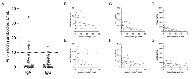 Figure 1
Peer Reviews
No public reviews on file for this paper yet. If you reviewed it on a platform where reviews are public (OpenReview, ICLR, NeurIPS, ICML), you can paste yours below so the community can read it here.
Videos
No videos yet. Explain this paper in a talk, walkthrough, or lecture? Add one.
Taxonomy
TopicsToxin Mechanisms and Immunotoxins · Pharmacological Effects of Natural Compounds · Transgenic Plants and Applications
Dear Editor,
Alpha (α)-fodrin, a non-erythroid homolog of spectrin, plays a role in maintaining the structural stability of cell membranes. During apoptosis, α-fodrin undergoes enzymatic cleavage by caspase-3, generating smaller fragments that act as neoantigens.^[1]^ Anti-α-fodrin antibodies are found in autoimmune disorders, including primary Sjögren’s syndrome (70%) and systemic lupus erythematosus (SLE; 10%–30%), as well as in a minority of healthy individuals (~2%).^[2,3]^ Experimental studies in animal models have demonstrated that immunization with α-fodrin induces lympho-cytic infiltration in salivary glands, while immunization with human Ro antigen results in the production of anti-α-fodrin antibodies, suggesting a potential shared intermolecular epitope between Ro and α-fodrin.^[4,5]^ However, the association between anti-α-fodrin antibodies and SLE-related clinical or immunological manifestations remains insufficiently investigated and is often overlooked in studies where SLE patients serve only as controls.
Our study aimed to elucidate the association between anti-α-fodrin antibodies and clinical as well as immunological perturbations in SLE, particularly their relationship with anti-Ro/anti-Ro52 antibodies and chemokine levels.
The study was approved by the local ethics committee (protocol number 16–960) and participants provided written informed consent. Thirty adult SLE patients (86% female; median age: 41 [19–63] years) without sicca symptoms, xero-stomia, xerophthalmia, or Sjögren’s syndrome participated in the study. Disease activity was assessed using the systemic lupus erythematosus disease activity index-2000 (SLEDAI-2K) score, with organ involvement categorized based on predefined criteria detailed elsewhere.^[6]^ Anti-α-fodrin IgA and IgG antibodies (cut-off ≥10 U/mL), as well as anti-Ro and an-ti-Ro52 antibodies (≥25 U/mL) (Orgentec; Mainz, Germany), were measured by Enzyme linked immunosorbent assay (ELISA). Positivity for anti-α-fodrin antibodies was defined by the detection of either IgA or IgG antibodies. Serum chemo-kines were measured using multiplexed bead-based assays (ThermoFisher Scientific; Minneapolis, United States).
Six patients tested positive for anti-α-fodrin IgA antibodies, while two tested positive for anti-α-fodrin IgG antibodies. Table 1 summarizes the main clinical and laboratory findings. No significant differences in demographics, comorbidities, disease severity, organ involvement, or medication usage were observed between antibody-positive and antibody-negative patients. Similarly, there were no differences in serum positivity for other SLE-associated antibodies. Median levels of anti-Ro (0, 3–35 U/mL vs. 0, 0–108 U/mL; P = 0.47) and anti-Ro52 (0, 0–21 U/mL vs. 0, 0–97 U/mL; P = 0.86) antibodies were similar between the groups.
The median levels of anti-α-fodrin IgA antibodies were 2.8 (0–34.3) U/mL, and for IgG antibodies, the median was 2.2 (0–14.0) U/mL (Figure 1–A). Disease activity analysis indicated that 26% of patients had quiescent disease, 66% had mild to moderate activity, and 6% had high activity. Nonetheless, no correlation was observed between SLEDAI-2K scores and anti-α-fodrin IgA (Spearman’s rho-0.02 [-0.39 to 0.34]; P = 0.89. Figure 1–B) or IgG (rho-0.04 [-0.40 to 0.33]; P = 0.81. Figure 1–E) antibody titers.
Chemokine analysis revealed that anti-α-fodrin-positive patients exhibited lower levels of C-C motif ligand 11 (CCL11)/eotaxin (54, 30–132 ng/L vs. 91, 23–339 ng/L; P = 0.02) and C-C motif ligand 2 (CCL2)/monocyte chemoattractant protein 1 (MCP-1) (136, 36–394 vs. 379, 107–2211 ng/L; P < 0.01) compared to antibody-negative patients. Other chemokine levels were similar between the groups (Table 1). Furthermore, levels of anti-α-fodrin immunoglobulin (IgA) antibodies were inversely correlated with CCL11/eotaxin (rho-0.38 [-0.66 to-0.02]; P = 0.03. Figure 1–C) and CCL2/MCP-1 (rho-0.39 [-0.66 to-0.03]; P = 0.02. Figure 1–D). Conversely, anti-α-fodrin IgG antibodies showed no significant correlation with CCL11/eotaxin (rho-0.32 [-0.62 to 0.04]; P = 0.07. Figure 1–F) or CCL2/MCP-1 (rho-0.03 [-0.39 to 0.34]; P = 0.86. Figure 1–G).
This study provides novel insights into the clinical and immunological roles of anti-α-fodrin antibodies in SLE. Contrary to initial expectations, no significant associations were observed between anti-α-fodrin antibodies and disease severity, organ involvement, or anti-Ro/anti-Ro52 antibodies in SLE patients. These findings contrast with animal models of Sjögren’s syndrome, where a connection between anti-α-fodrin and anti-Ro antibodies has been suggested.^[4,5,7]^ Interestingly, our findings reveal a potential link between anti-α-fodrin antibodies and reduced levels of certain chemokines, suggesting a possible protective role in modulating the inflammatory response triggered by α-fodrin-induced apoptosis. This mechanism aligns with other cases of antibody-mediated regulation of harmful proteins in SLE, such as anti-cytokine autoantibodies that neutralize excessive cytokine activity and mitigate tissue damage.^[8]^ A plausible mechanism is that anti-α-fodrin antibodies may sequester free α-fodrin, preventing its activation of caspase-3 and subsequent apoptosis. This could reduce the production of chemokines like CCL2/MCP-1 and CCL11/eotaxin, which are crucial for recruiting immune cells such as monocytes and granulocytes.^[9,10]^
Although the small sample size and cross-sectional design limit the ability to draw definitive conclusions, our study failed to establish a clear association between anti-α-fodrin antibodies and anti-Ro/anti-Ro52 antibodies, disease severity, or organ-specific involvement in SLE. Nevertheless, our findings suggest a potential mechanistic role for these antibodies in regulating chemokine-driven inflammation.
The reference list from the paper itself. Each links out to its DOI / PubMed record.
- 1Nellikka RK Sreeja JS Dharmapal Dα-Fodrin is required for the organization of functional microtubules during mitosis Cell Cycle 201918271327263145518610.1080/15384101.2019.1656476 PMC 6773225 · doi ↗ · pubmed ↗
- 2Shen L Suresh L Autoantibodies, detection methods and panels for diagnosis of Sjögren’s syndrome Clin Immunol 201718224292839096510.1016/j.clim.2017.03.017 · doi ↗ · pubmed ↗
- 3Hu Q Wang D Chen W The accuracy of the anti-α-fodrin antibody test for diagnosis of Sjögren’s syndrome: a meta-analysis Clin Biochem 201346137213762363967610.1016/j.clinbiochem.2013.04.020 · doi ↗ · pubmed ↗
- 4Jing He Hui Wang Jin-xia Zhao[Establishment of Sjögren’s syndrome models by immunization with alpha-Fodrin: experiment with mice]Zhonghua Yi Xue Za Zhi 2008882360236319087701 · pubmed ↗
- 5Kurien BT Dorri Y Bachmann M Scofield RH Induction of anti-Ro 60/anti-La by immunisation with spectrin and induction of anti-spectrin by immunisation with Ro 60 and 4-hydroxy-2-nonenal-modified Ro 60 immunisation Clin Exp Rheumatol 20123088689322776429 PMC 5664947 · pubmed ↗
- 6Amezcua-Guerra LMMárquez-Velasco R Chávez-Rueda AK Type III Interferons in Systemic Lupus Erythematosus: Association Between Interferon λ3, Disease Activity, and Anti-Ro/SSA Antibodies J Clin Rheumatol 2017233683752893747210.1097/RHU.0000000000000581 · doi ↗ · pubmed ↗
- 7Ding M Zhang J Epitope spreading induced by immunization with synthetic SSB peptides Exp Ther Med 2016121471502734703010.3892/etm.2016.3267 PMC 4906616 · doi ↗ · pubmed ↗
- 8Howe HS Leung BPL Anti-Cytokine Autoantibodies in Systemic Lupus Erythematosus Cells 20199723189220010.3390/cells 9010072 PMC 7016754 · doi ↗ · pubmed ↗