Unusual Presentation of Epidermodysplasia Verruciformis (EV) in Non-Sun Exposed Area: A Case Report
Nidal Jebrini, Majed Dwaik, Mohanad Jaber, Sami Jabari, Raghad Razem, Man Sarahna, Rashad Alzaro, Feras Aljabari, Mohamed Aqel, Husein Sarahneh

TL;DR
A rare case of skin cancer in a sun-protected area was found in a woman with a genetic skin condition called EV, challenging the usual understanding of where such cancers occur.
Contribution
This is the second global case of SCC in a non-sun-exposed area among EV patients, challenging the sun-exposure paradigm.
Findings
A 28-year-old woman with EV developed SCC on her scalp, a non-sun-exposed area.
The patient had multiple lesions resembling trichoblastic and verrucous carcinoma.
This case highlights the need for close monitoring and UV protection in EV patients.
Abstract
Introduction and Importance: Epidermodysplasia verruciformis (EV), a rare hereditary skin disorder linked to HPV immunity, increases the risk of squamous cell carcinoma (SCC), typically in sun-exposed areas. This case highlights an extraordinary instance of SCC in a Sun-shielded region, marking the second documented case globally. Methods: The medical records and histopathological slides of the case were retrospectively reviewed. This work has been reported based on the CARE criteria. Case Presentation: A 28-year-old Palestinian woman, who adheres to a sun-protective Hijab due to her Muslim faith and has limited sun exposure working in a clothing store, with painful scalp lesions presented at the dermatology clinic. She and her siblings were diagnosed with EV. Three years ago, a painful, enlarging lesion on her scalp led to a diagnosis of trichoblastic carcinoma, followed by the…
Genes, proteins, chemicals, diseases, species, mutations and cell lines named across the full text — each resolved to its canonical identifier and authoritative record.
Click any figure to enlarge with its caption.
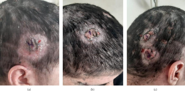 Figure 1
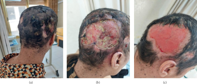 Figure 2
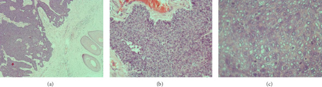 Figure 3
Peer Reviews
No public reviews on file for this paper yet. If you reviewed it on a platform where reviews are public (OpenReview, ICLR, NeurIPS, ICML), you can paste yours below so the community can read it here.
Videos
No videos yet. Explain this paper in a talk, walkthrough, or lecture? Add one.
Taxonomy
TopicsGenetic and rare skin diseases. · Cancer and Skin Lesions · Hedgehog Signaling Pathway Studies
Summary
- • The occurrence of epidermodysplasia verruciformis (EV) in sun-shielded areas is exceptionally rare, with few exceptions noted in this case study.
- • EV significantly heightens susceptibility to nonmelanoma skin cancer (NMSC), primarily squamous cell carcinoma (SCC).
- • Early lesion identification and prompt retinoid therapy are crucial in impeding disease progression and reducing recurrence risk.
1. Introduction
EV is an exceptionally rare and lifelong hereditary dermatological condition that commonly presents during infancy or early childhood. This disorder disrupts the body's immune defense mechanisms, rendering it incapable of effectively guarding against specific varieties of human papillomavirus (HPV), particularly those belonging to the beta-HPV subgroup. Consequently, individuals afflicted with EV exhibit a distinctive combination of plane warts and lesions resembling pityriasis versicolor from an early age [1].
Patients with EV face a heightened susceptibility to the development of SCC, particularly in their third and fourth decades of life. Notably, these malignancies tend to manifest predominantly in sun-exposed regions of the skin, underscoring the pivotal role of ultraviolet radiation as a significant cocarcinogenic factor, in conjunction with HPV infection [2]. Within the context of EV-associated carcinomas, HPV DNA sequences, most notably HPV Types 5 and 8, consistently feature as identified pathogens within these neoplastic lesions [3]. Here, we present a rare case of a patient with EV who has developed SCC in a non–sun-exposed area of the skin.
2. Case Presentation
A 28-year-old Palestinian woman presented to the dermatology clinic with multiple painful, ulcerative, and purulent lesions scattered across her scalp. The lesions had developed gradually over the past year. Due to socioeconomic challenges, she had delayed seeking medical attention. As part of her religious practice, she habitually covers her scalp with a Hijab, limiting sun exposure. Additionally, her work in a clothing store confines her to an indoor environment, contributing to minimal sun exposure. However, while limited sunlight may be relevant to her condition, it is not assumed to be a primary factor in lesion development.
The patient has a notable history of EV, diagnosed at age 13 when she and her siblings presented with wart-like lesions on the head, face, and upper extremities. To manage EV-associated inflammation, she was started on isotretinoin therapy, although details of this treatment's initiation and duration were not closely documented. Prior to her current presentation, she had discontinued isotretinoin. While this discontinuation was temporally followed by an increase in scalp lesions, isotretinoin's therapeutic effect is limited and does not offer definitive prevention against malignant transformation in EV patients.
Approximately 3 years before this presentation, one lesion on her scalp began enlarging and became painful, prompting medical evaluation. Following an initial dermatology assessment, she was referred to oncology due to suspicion of malignancy. Surgical excision of the lesion was performed by a plastic surgeon, with histopathology confirming trichoblastic carcinoma (Figure 1). Over the next several months, six additional lesions with similar characteristics appeared on her scalp, requiring further surgical intervention.
One year later, the patient returned to the clinic with increasing numbers of painful, pus-producing scalp lesions, primarily located in the right occipital area. Complete excision of a 6 × 9-cm region of the right occipital scalp was undertaken (Figure 2), with the specimens sent for the histopathological analysis (Figure 3). The findings revealed both trichoblastic carcinoma and verrucous carcinoma. Although scalp localization is consistent with known patterns for trichoblastic lesions, the connection between EV and lesion localization on the scalp remains speculative, given the multifactorial nature of these conditions and the need for further research.
3. Discussion
EV is an exceptionally rare genetic skin disorder with an autosomal recessive inheritance pattern, affecting less than one in every 1,000,000 individuals. Typically, it manifests during childhood, presenting as widespread flat warts resembling pityriasis, with an average onset age of 4.9 years [4].
The diagnosis of EV relies on clinical, histopathological, and molecular findings. Clinical features, such as the failure to eliminate lesions after therapy, and histopathological characteristics, such as gray–blue cytoplasm and enlarged nuclei, contribute to the diagnosis. Autosomal recessive abnormalities in EVER1/TCM6 or EVER2/TCM8 may cause classical hereditary EV, with these genes located on Chromosome 17 [5–7].
EV is strongly correlated with HPV, particularly beta-HPV Types 5, 8, 9, 12, 14, 15, and 17 and Types 19–25, 36–38, 47, and 50. High-risk strains such as HPV-5 and HPV-8 significantly increase the risk of NMSC [6, 8]. Mutations in TMC genes increase susceptibility to specific HPV infections, predisposing individuals to EV [8, 9]. Cutaneous malignancies occur in 30%–70% of EV cases, typically after age 30, with SCC manifesting in sun-exposed areas [10]. Our case deviates from the norm, presenting SCC in non–sun-exposed skin, a rarity with only one similar case reported [5, 6].
Our case mirrors a unique instance in a 25-year-old Iranian male, challenging the notion that SCC in EV primarily arises in sun-exposed areas. Both cases emphasized the importance of monitoring individuals with EV, irrespective of sun exposure, showcasing clinical diversity [5].
The treatment for EV centers on preventing benign lesions from progressing to malignancy. UV protection from early childhood and oral retinoids are recommended [11]. It is essential to note that the effects of retinoid therapy are often reversible upon discontinuation. These medications offer several benefits, including antiviral properties and the regulation of epithelial cell differentiation, thereby assisting in the management of disease progression [4, 12]. In our case, the patient discontinued isotretinoin, leading to lesion recurrence and necessitating multiple surgical interventions, including a 6 × 9-cm scalp excision to proactively prevent further recurrence.
4. Patient's Persecriptive
The patient's perspective provides a firsthand account of the challenges they have faced due to lesions on their scalp, which have gradually emerged over the past year, leading to considerable physical and emotional distress. Despite adhering to religious practices necessitating scalp coverage and predominantly working indoors, the persistence of these lesions has significantly disrupted their daily life.
Furthermore, the patient's account of a familial history involving similar lesions, initially diagnosed as EV, suggests a predisposition to cutaneous malignancies. This raises concerns regarding disease progression and complicating factors such as trichoblastic carcinoma and verrucous carcinoma, as revealed through the recent surgical intervention.
In the patient's own words: “The pain got worse, especially at night, and these lesions produce a lot of pus. Recently, they did a big surgery and found both trichoblastic carcinoma and verrucous carcinoma. Doctors explained that “It's a tough situation, but I'm hoping they'll find a way to help me feel better. Just want this pain and uncertainty to go away.”
Given the patient's poignant narrative and the complexity of their medical journey, it is imperative to delve into the long-term outcomes following surgical interventions for SCC on the scalp. This entails a thorough assessment of postoperative complications, including wound healing issues, infection, and the potential for SCC recurrence. Additionally, developing comprehensive strategies for pain management and addressing the patient's uncertainties regarding their prognosis are essential for providing holistic care and alleviating their distress.
5. Conclusion
In this particular case, we present an exceedingly rare occurrence wherein a patient diagnosed with EV has developed SCC in a region of the skin that is not typically exposed to sunlight. The rarity of this case underscores its exceptional nature within the context of EV-associated complications.
The reference list from the paper itself. Each links out to its DOI / PubMed record.
- 1Lewandowsky F. Lutz W. Ein Fall Einer Bisher Nicht Beschriebenen Hauterkrankung (Epidermodysplasia Verruciformis) Archiv für Dermatologie und Syphilis 1922141219320310.1007/bf 019388332-s 2.0-0001389413 · doi ↗
- 2Sullivan M. Ellis F. A. Epidermodysplasia Verruciformis (Lewandowsky and Lutz) Archives of Dermatology and Syphilology 1939403422432
- 3Ramoz N. Favre M. Orth G. Evidence for a Nonallelic Heterogeneity of Epidermodysplasia Verruciformis With Two Susceptibility Loci Mapped to Chromosome Regions 2p 21–P 24 and 17q 25 Journal of Investigative Dermatology 200011461148115310.1046/j.1523-1747.2000.00996.x 2-s 2.0-003412411210844558 · doi ↗ · pubmed ↗
- 4Burger B. Itin P. Epidermodysplasia Verruciformis Current Problems in Dermatology 20144512313110.1159/0003560682-s 2.0-8492248022924643182 · doi ↗ · pubmed ↗
- 5Ansarin H. Tajziehchi L. Shaianfar N. A Case of Epidermodysplasia Verruciformis With Squamous Cell Carcinomas on Non-Sun-Exposed Areas of Skin Archives of Iranian Medicine 200710226126317367237 · pubmed ↗
- 6Berk D. R. Bruckner A. L. Lu D. Epidermodysplasia Verruciform-Like Lesions in an HIV Patient Dermatology Online Journal 2009151 p. 110.5070/d 36ks 5t 6qw 19281706 · doi ↗ · pubmed ↗
- 7Ramoz N. Rueda L. A. Bouadjar B. Montoya L. S. Orth G. Favre M. Mutations in Two Adjacent Novel Genes Are Associated With Epidermodysplasia Verruciformis Nature Genetics 200232457958110.1038/ng 10442-s 2.0-1874436988612426567 · doi ↗ · pubmed ↗
- 8Oiso N. Kubo A. Shimizu A. Epidermodysplasia Verruciformis Without Progression to Squamous Cell Carcinomas in an Elderly Man: α-Human Papillomavirus Infection in the Evolving Verruca International Journal of Dermatology 2020599 e 334e 33610.1111/ijd.1488332406058 · doi ↗ · pubmed ↗