Three-dimensional exploration of the chicken embryo, a comparative study of light sheet and histological visualisation
M. W. Smallridge, T. E. Aktepe, M. J. C. Coppo, P. K. Vaz, A. Diaz-Méndez, C. M. Murray, G. Segal, J. M. Devlin, C. A. Hartley

TL;DR
This study compares light sheet microscopy and traditional histology to visualize chicken embryos in 3D, enhancing anatomical understanding.
Contribution
The study introduces an ethyl cinnamate-based tissue clearing method for light sheet imaging of chicken embryos.
Findings
Light sheet imaging provided clear 3D visualization of organ structures using autofluorescence.
Combining histology with light sheet imaging enhanced anatomical mapping and contextual insights.
Volumetric projections of lung vasculature and heart connections were successfully rendered.
Abstract
Ultramicroscopy has offered new avenues into the visualisation of tissues within animal models, providing three-dimensional visualisation through the use of light sheet fluorescence microscopy. This study aimed to develop and apply an optical tissue clearing method to investigate the application of light sheet fluorescence microscopy to image late-stage chicken embryos, and compare anatomical visualisation to traditional histological staining. Seventeen-day old specific pathogen free embryos were collected, fixed, and sectioned. Haematoxylin and eosin stained sections were prepared for histology, while light sheet imaging required the tissues to be optically clear. For this, an ethyl cinnamate-based method was utilised, allowing for acquisition of clear, unobstructed three-dimensional images of significant organ structures and systems using only autofluorescence. The use of established…
Genes, proteins, chemicals, diseases, species, mutations and cell lines named across the full text — each resolved to its canonical identifier and authoritative record.
Click any figure to enlarge with its caption.
 Figure 1
 Figure 2
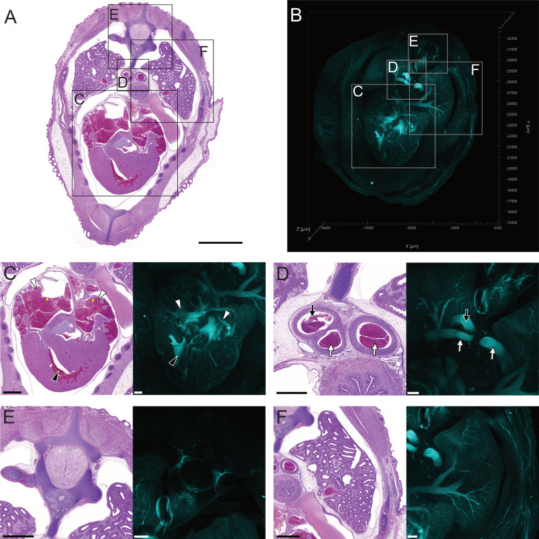 Figure 3
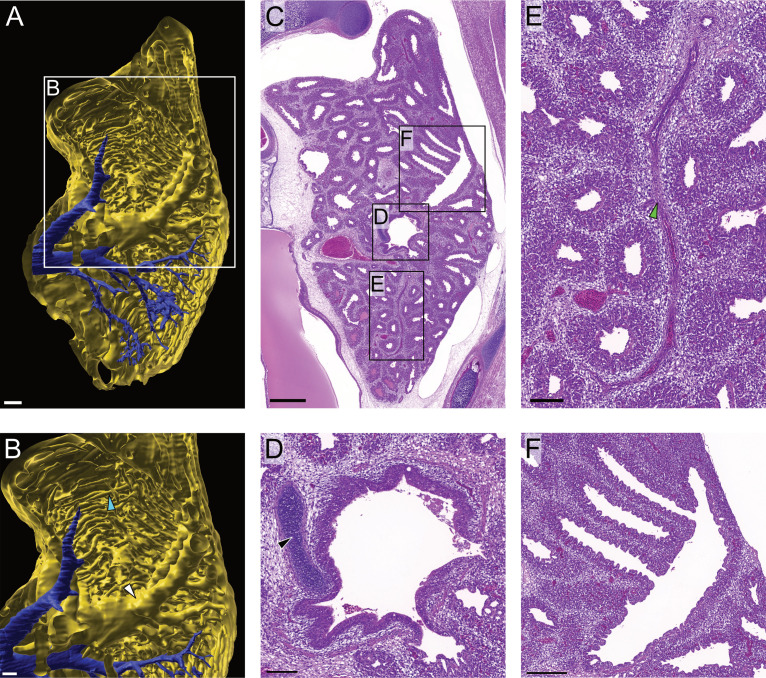 Figure 4
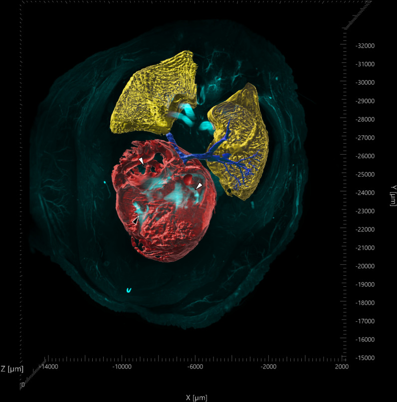 Figure 5
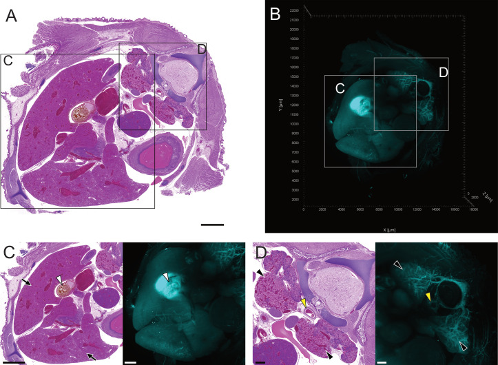 Figure 6
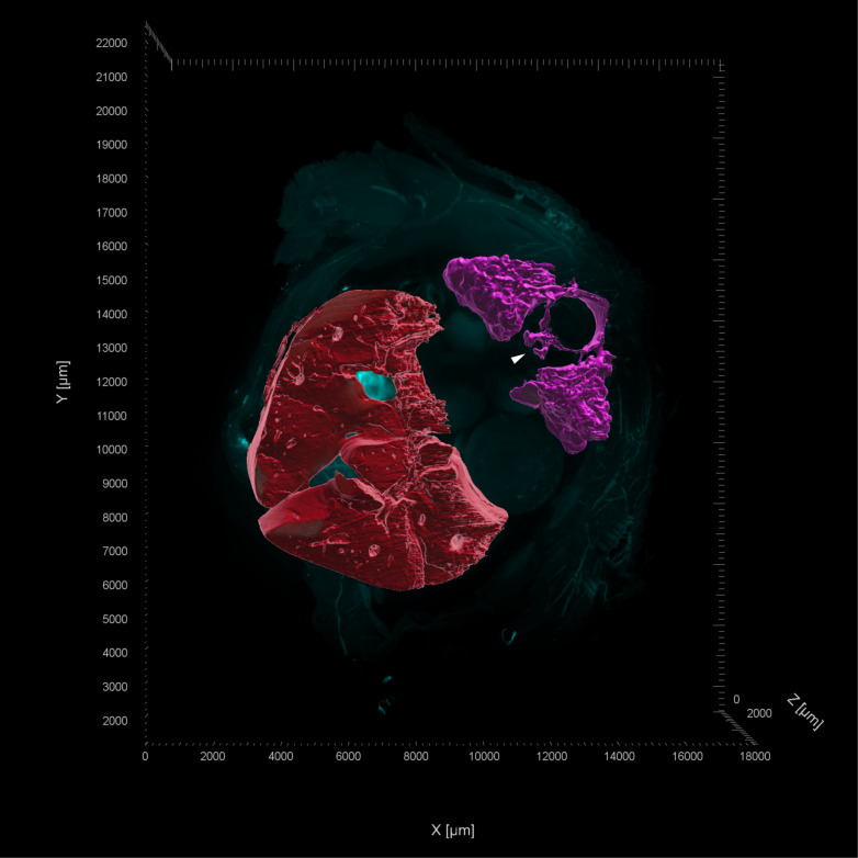 Figure 7
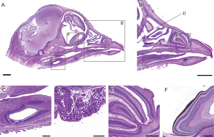 Figure 8
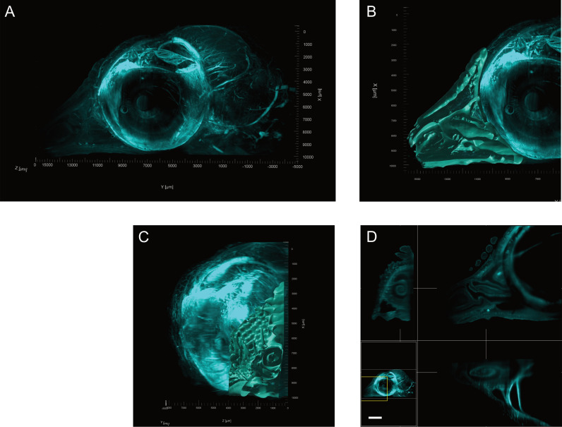 Figure 9
Peer Reviews
No public reviews on file for this paper yet. If you reviewed it on a platform where reviews are public (OpenReview, ICLR, NeurIPS, ICML), you can paste yours below so the community can read it here.
Videos
No videos yet. Explain this paper in a talk, walkthrough, or lecture? Add one.
Taxonomy
TopicsAdvanced Fluorescence Microscopy Techniques · Cell Image Analysis Techniques · Single-cell and spatial transcriptomics