Complication rates in real-time ultrasound-guided vs static echocardiography-guided pericardiocentesis: a cohort study
Virginia Zarama, Carlos E. Vesga, John Balanta-Silva, Mario M. Barbosa, Jaime A. Quintero, Ana Clarete, Paula A. Vesga-Reyes, Juan Carlos Silva Godinez

TL;DR
This study compares complication rates of two pericardiocentesis techniques and finds both are similarly safe, with real-time ultrasound showing potential benefits.
Contribution
The study evaluates the safety of real-time ultrasound-guided pericardiocentesis compared to the traditional static echocardiography-guided method.
Findings
Real-time in-plane US-guided pericardiocentesis had a 97% success rate compared to 93% for static echo-guided procedures.
Only one major complication occurred with the real-time technique versus four with the static approach.
Total complication rates were not significantly different between the two techniques.
Abstract
Static echocardiography-guided pericardiocentesis, the current standard of care, uses a phased-array probe to locate the largest fluid pocket, marking the safest entry site and needle trajectory. Nevertheless, real-time needle visualization throughout the procedure would potentially increase success and decrease complications. The aim of this study was to assess the complication rates of the real-time in-plane ultrasound-guided technique compared to the traditional static echocardiography-guided pericardiocentesis. All adult patients who underwent pericardiocentesis in a tertiary care hospital from January 2011 to June 2024 were identified. The incidence of total complications of the real-time, in-plane, US-guided pericardiocentesis versus the static echocardiography-guided technique was compared using a regression model with overlap weighting, based on propensity scores, to adjust for…
Genes, proteins, chemicals, diseases, species, mutations and cell lines named across the full text — each resolved to its canonical identifier and authoritative record.
Click any figure to enlarge with its caption.
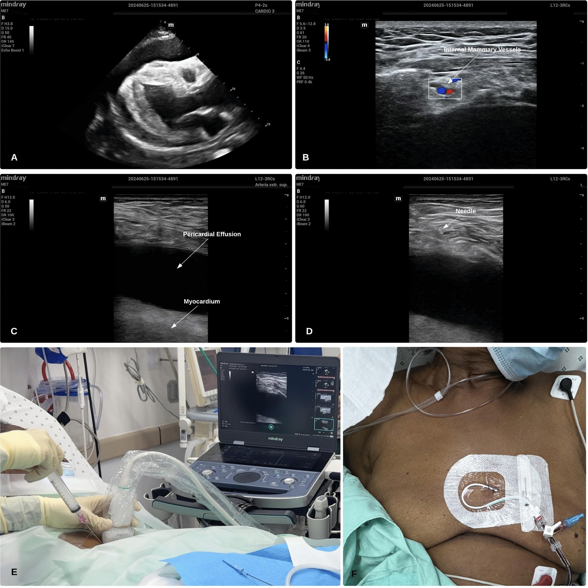 Figure 1
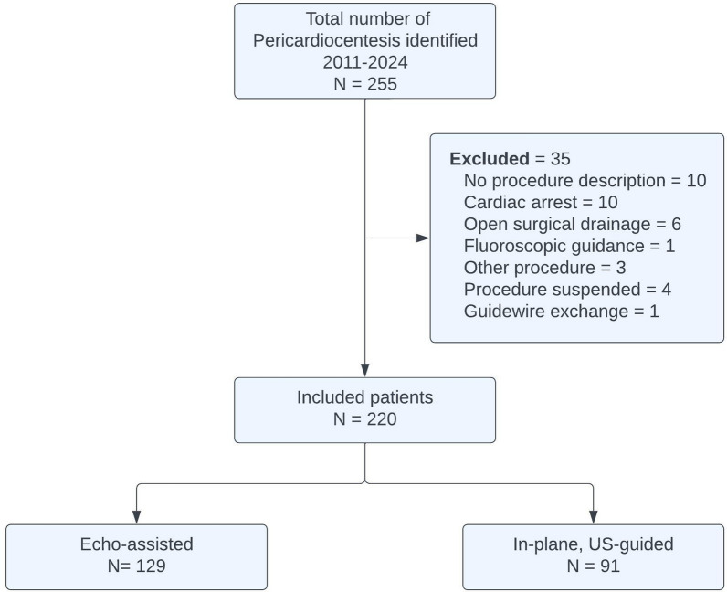 Figure 2
 Figure 3
 Figure 4
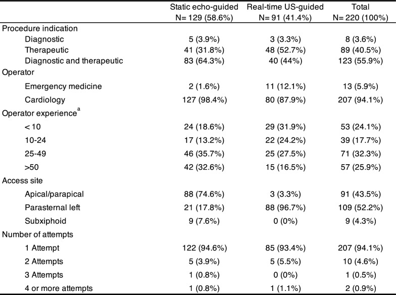 Figure 5
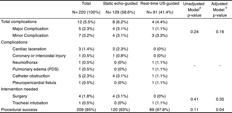 Figure 6
Peer Reviews
No public reviews on file for this paper yet. If you reviewed it on a platform where reviews are public (OpenReview, ICLR, NeurIPS, ICML), you can paste yours below so the community can read it here.
Videos
No videos yet. Explain this paper in a talk, walkthrough, or lecture? Add one.
Taxonomy
TopicsUltrasound in Clinical Applications · Pericarditis and Cardiac Tamponade · Hemodynamic Monitoring and Therapy