Feasibility of optimal vertex size and spacing for lattice radiotherapy implementation using helical tomotherapy
Yunji Seol, Young Kyu Lee, Byeong Jin Kim, Kyu Hye Choi, Ji Hyun Hong, Chan-beom Park, Sun Hwa Kim, Hyeong Wook Park, Jung-Il Kim, Wonjoong Cheon, Young-nam Kang, Byung Ock Choi

TL;DR
This study explores how vertex size and spacing affect dose distribution in lattice radiotherapy using helical tomotherapy, aiming to optimize treatment effectiveness.
Contribution
The study quantitatively evaluates the feasibility of lattice radiotherapy using helical tomotherapy and identifies optimal vertex size and spacing for achieving desired dose ratios.
Findings
VPDR decreased significantly with increasing vertex size and spacing.
The SI direction consistently showed lower VPDR values compared to AP and LAT directions.
Smaller vertex sizes required larger spacing to achieve VPDR values below 0.4.
Abstract
Lattice radiotherapy (LRT), a type of spatially fractionated radiotherapy (SFRT), delivers high dose at specific volumes of lattice structure within the tumor to create a low valley-to-peak dose ratio (VPDR). This study aims to evaluate the feasibility of implementing SFRT using helical tomotherapy and to investigate the effects of vertex size and spacing for attaining the VPDR. A three-dimensional lattice structure with 3×3×3 vertices was designed in a cheese phantom. Vertex sizes of 0.5 cm, 1.0 cm, and 2.0 cm were assessed, with spacing from 1.0 cm to 5.0 cm. The prescribed dose was set to 20 Gy to the vertices in a single fraction. VPDR was calculated from dose profiles along lines connecting three vertices in the anterior-posterior (AP), lateral (LAT), and superior-inferior (SI) directions. The minimum, maximum, and mean dose for each vertex, as well as conformity, homogeneity and…
Genes, proteins, chemicals, diseases, species, mutations and cell lines named across the full text — each resolved to its canonical identifier and authoritative record.
Click any figure to enlarge with its caption.
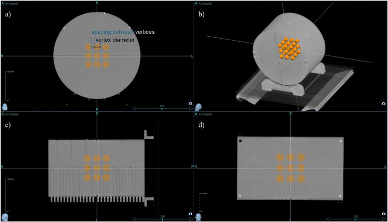 Figure 1
 Figure 2
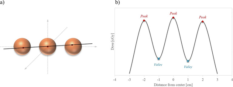 Figure 3
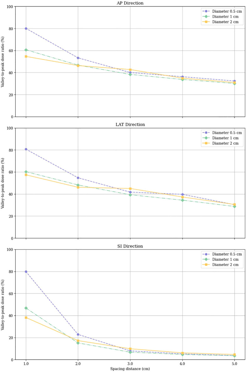 Figure 4
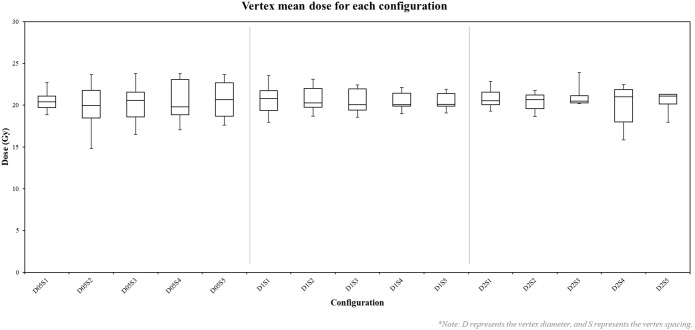 Figure 5
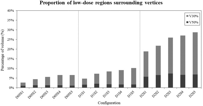 Figure 6
Peer Reviews
No public reviews on file for this paper yet. If you reviewed it on a platform where reviews are public (OpenReview, ICLR, NeurIPS, ICML), you can paste yours below so the community can read it here.
Videos
No videos yet. Explain this paper in a talk, walkthrough, or lecture? Add one.
Taxonomy
TopicsAdvanced Radiotherapy Techniques · Radiation Detection and Scintillator Technologies · Brain Metastases and Treatment