Impact of contralateral pelvic drop and femoral adduction on the femoral head acetabular coverage: A study on the reproducibility of a new radiographic measurement method
Renato Locks, Eliane C. Guadagnin, Guilherme Pradi Adam, Felipe F. Gonzalez, Jorge Chahla, Liszt Palmeira de Oliveira, Leonardo Metsavaht, Gustavo Leporace

TL;DR
This study shows that a new radiographic method incorporating motion during running can reliably assess hip coverage, improving understanding for active individuals.
Contribution
The study introduces a reproducible radiographic method that integrates dynamic motion for assessing acetabular coverage.
Findings
Most radiographic measurements showed good to excellent inter- and intra-rater reliability.
Dynamic motion analysis combined with radiography could enhance surgical decision-making for hip conditions.
Contralateral pelvic drop and femoral adduction during running significantly affect acetabular coverage measurements.
Abstract
Traditional radiographic measurements for acetabular dysplasia and femoroacetabular impingement syndrome (FAIS) are typically done in static positions, overlooking dynamic behaviours. This study investigated the reproducibility of a new radiographic method that incorporates pelvic and femoral motion during running. This cross‐sectional retrospective study included 10 patients (5 males/5 females; Mean 42.4, SD: 3.0 years) with symptomatic unilateral FAIS. Participants underwent three‐dimensional running analysis and standard supine anteroposterior (AP) pelvis radiographs. Using specialised software, the femur and pelvis were rotated in the coronal plane according to peak angles of contralateral pelvic drop and femoral adduction from the running analysis, preserving the original hip joint centre. Two experienced physicians measured the lateral centre edge angle (LCEA), acetabular index…
Genes, proteins, chemicals, diseases, species, mutations and cell lines named across the full text — each resolved to its canonical identifier and authoritative record.
Click any figure to enlarge with its caption.
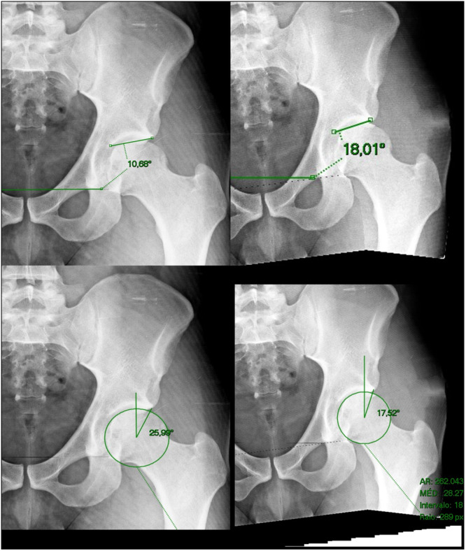 Figure 1
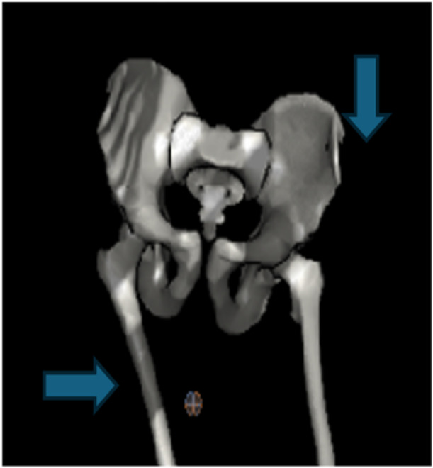 Figure 2
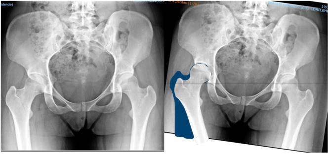 Figure 3
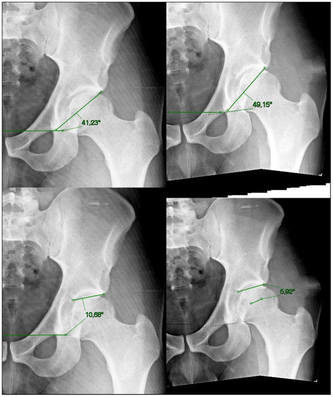 Figure 4
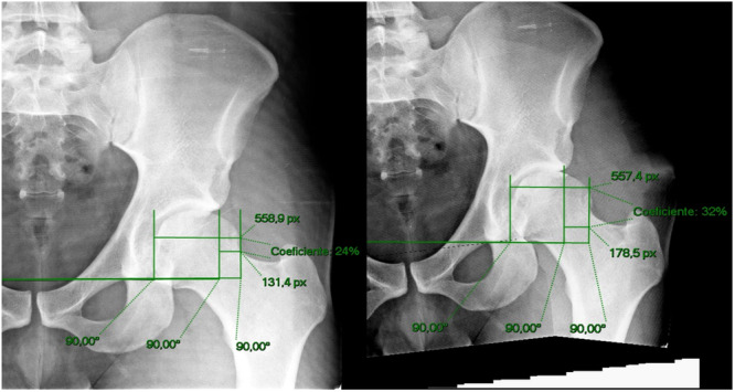 Figure 5
Peer Reviews
No public reviews on file for this paper yet. If you reviewed it on a platform where reviews are public (OpenReview, ICLR, NeurIPS, ICML), you can paste yours below so the community can read it here.
Videos
No videos yet. Explain this paper in a talk, walkthrough, or lecture? Add one.
Taxonomy
TopicsHip disorders and treatments · Lower Extremity Biomechanics and Pathologies · Bone and Joint Diseases