The genome sequence of the common crane, Grus grus (Linnaeus, 1758)
Michelle F. O’Brien, Rosa Lopez Colom, Bengt Hansson, Annabel Whibley

TL;DR
This paper presents the genome sequence of the common crane, including detailed assemblies of its haplotypes and mitochondrial genome.
Contribution
The study provides a high-quality genome assembly for Grus grus, including two haplotypes and the mitochondrial genome.
Findings
The genome assembly includes two haplotypes with lengths of 1,352.26 and 1,291.08 megabases.
Haplotype 1 is scaffolded into 40 chromosomal pseudomolecules, including the Z sex chromosome.
The mitochondrial genome is assembled and is 18.9 kilobases in length.
Abstract
We present a genome assembly from a male specimen of Grus grus (common crane; Chordata; Aves; Gruiformes; Gruidae). The assembly contains two haplotypes with total lengths of 1,352.26 megabases and 1,291.08 megabases. Most of haplotype 1 (91.85%) is scaffolded into 40 chromosomal pseudomolecules, including the Z sex chromosome. Haplotype 2 was assembled to scaffold level. The mitochondrial genome has also been assembled and is 18.9 kilobases in length.
Genes, proteins, chemicals, diseases, species, mutations and cell lines named across the full text — each resolved to its canonical identifier and authoritative record.
Click any figure to enlarge with its caption.
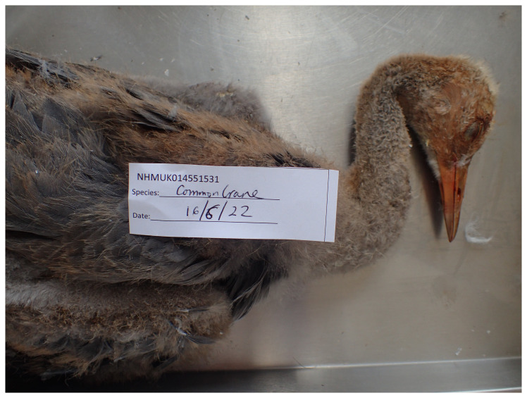 Figure 1
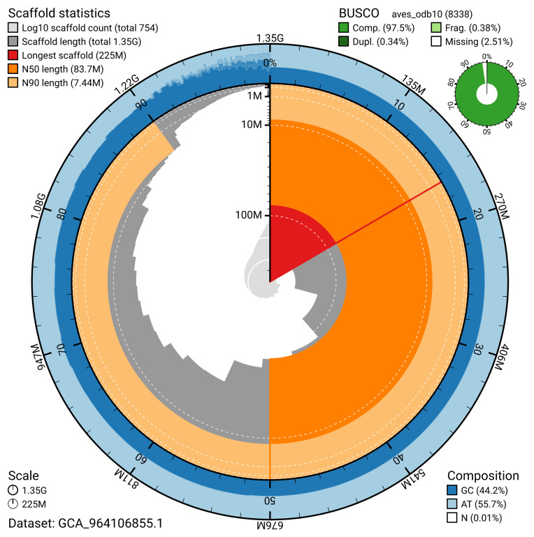 Figure 2
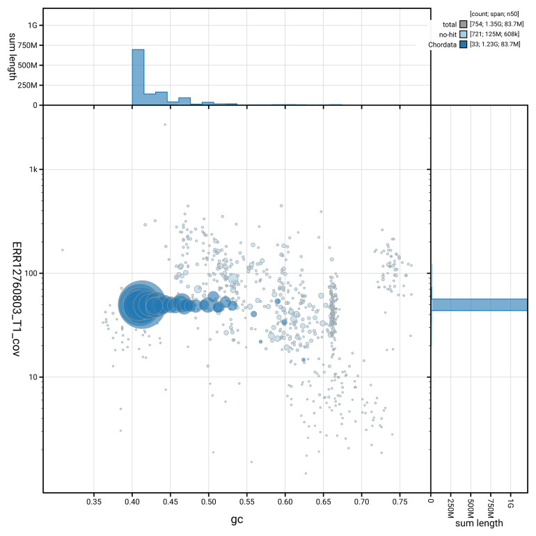 Figure 3
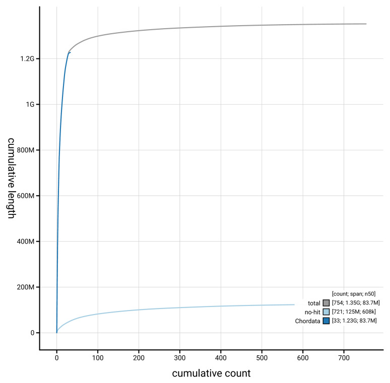 Figure 4
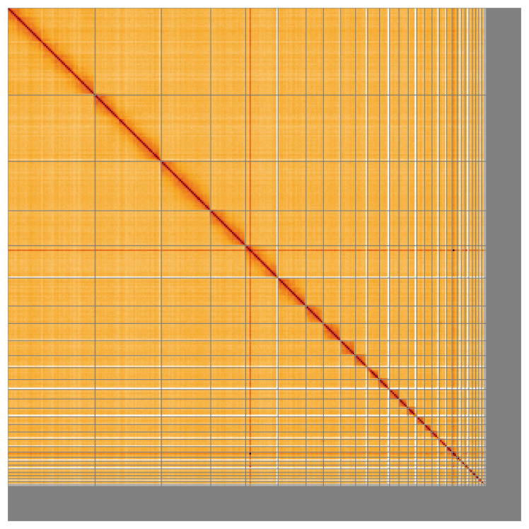 Figure 5
| Project information | |||
|---|---|---|---|
|
| Grus grus (Eurasian crane) | ||
|
| PRJEB73916 | ||
|
|
| ||
|
| SAMEA113398837 | ||
|
| 40816 | ||
| Specimen information | |||
|
|
|
|
|
|
| bGruGru1 | SAMEA113398891 | muscle |
|
| bGruGru1 | SAMEA113398891 | muscle |
|
| bGruGru1 | SAMEA113398891 | muscle |
| Sequencing information | |||
|
|
|
|
|
|
| ERR12765181 | 3.80e+08 | 57.45 |
|
| ERR12760803 | 6.94e+06 | 63.3 |
|
| ERR13493931 | 9.30e+07 | 14.04 |
| Genome assembly | Haplotype 1 | Haplotype 2 |
|---|---|---|
| Assembly name | bGruGru1.hap1.1 | bGruGru1.hap2.1 |
| Assembly accession | GCA_964106855.1 | GCA_964059345.1 |
| Assembly level | chromosome | scaffold |
| Span (Mb) | 1,352.26 | 1,291.08 |
| Number of contigs | 1,346 | 885 |
| Number of scaffolds | 753 | 326 |
| Longest scaffold (Mb) | 224.75 | None |
| Assembly metrics (benchmark) | Haplotype 1 | Haplotype 2 |
| Contig N50 length (≥ 1 Mb) | 3.37 Mb | 3.59 Mb |
| Scaffold N50 length (= chromosome N50) | 83.71 Mb | 84.07 Mb |
| Consensus quality (QV) (≥ 40) | 63.4 | 64.8 |
|
| 91.47% | 91.30% |
| Combined
| 99.73% | |
| BUSCO
| C:97.5%[S:97.2%,D:0.3%], F:0.4%,M:2.1%,n:8,338 | - |
| Percentage of assembly mapped to
| 91.85% | - |
| Sex chromosomes (localised homologous pairs) | Z | - |
| Organelles (one complete allele) | Mitochondrial genome: 18.9 kb | - |
| INSDC accession | Name | Length (Mb) | GC% |
|---|---|---|---|
| 1 | 224.75 | 41.5 | |
| 2 | 170.94 | 41 | |
| 3 | 127.3 | 41 | |
| 4 | 83.71 | 41 | |
| 5 | 72.02 | 42 | |
| 6 | 45.01 | 42.5 | |
| 7 | 44.52 | 43 | |
| 8 | 38.18 | 43 | |
| 9 | 31.19 | 44 | |
| 10 | 30.35 | 46.5 | |
| 11 | 25.8 | 44 | |
| 12 | 24.54 | 44 | |
| 13 | 23.83 | 43 | |
| 14 | 22.08 | 45 | |
| 15 | 19.77 | 45.5 | |
| 16 | 19.38 | 46.5 | |
| 17 | 19.17 | 47 | |
| 18 | 18.76 | 50 | |
| 19 | 13.8 | 47 | |
| 20 | 13.25 | 48.5 | |
| 21 | 10.41 | 47.5 | |
| 22 | 9.99 | 52 | |
| 23 | 9.62 | 51.5 | |
| 24 | 9.38 | 50.5 | |
| 25 | 8.32 | 49.5 | |
| 26 | 7.69 | 51 | |
| 27 | 7.44 | 53 | |
| 28 | 7.17 | 53 | |
| 29 | 2.17 | 61 | |
| 30 | 2.09 | 56 | |
| 31 | 1.7 | 60 | |
| 32 | 1.69 | 59 | |
| 33 | 1.67 | 61.5 | |
| 34 | 1.6 | 53 | |
| 35 | 1.51 | 55.5 | |
| 36 | 0.49 | 62.5 | |
| 37 | 0.4 | 63 | |
| 38 | 0.4 | 57 | |
| 39 | 0.21 | 65.5 | |
| Z | 89.82 | 41 | |
| MT | 0.02 | 44.5 |
| Software tool | Version | Source |
|---|---|---|
| BEDTools | 2.30.0 |
|
| BLAST | 2.14.0 |
|
| BlobToolKit | 4.3.9 |
|
| BUSCO | 5.5.0 |
|
| bwa-mem2 | 2.2.1 |
|
| Cooler | 0.8.11 |
|
| DIAMOND | 2.1.8 |
|
| fasta_windows | 0.2.4 |
|
| FastK | 427104ea91c78c3b8b8b49f1a7d6bbeaa869ba1c |
|
| Gfastats | 1.3.6 |
|
| GoaT CLI | 0.2.5 |
|
| Hifiasm | 0.19.8-r603 |
|
| HiGlass | 44086069ee7d4d3f6f3f0012569789ec138f42b84aa44357826c0b6753eb28de |
|
| Merqury.FK | d00d98157618f4e8d1a9190026b19b471055b22e |
|
| Minimap2 | 2.24-r1122 |
|
| MitoHiFi | 3 |
|
| MultiQC | 1.14, 1.17, and 1.18 |
|
| NCBI Datasets | 15.12.0 |
|
| Nextflow | 23.10.0 |
|
| PretextView | 0.2 |
|
| samtools | 1.19.2 |
|
| sanger-tol/ascc | - |
|
| sanger-tol/blobtoolkit | 0.5.1 |
|
| Seqtk | 1.3 |
|
| Singularity | 3.9.0 |
|
| TreeVal | 1.2.0 |
|
| YaHS | 1.2a.2 |
|
- —Wellcome Trust
Peer Reviews
No public reviews on file for this paper yet. If you reviewed it on a platform where reviews are public (OpenReview, ICLR, NeurIPS, ICML), you can paste yours below so the community can read it here.
Videos
No videos yet. Explain this paper in a talk, walkthrough, or lecture? Add one.
Taxonomy
TopicsGenomics and Phylogenetic Studies · Bat Biology and Ecology Studies · Virology and Viral Diseases
Species taxonomy
Eukaryota; Opisthokonta; Metazoa; Eumetazoa; Bilateria; Deuterostomia; Chordata; Craniata; Vertebrata; Gnathostomata; Teleostomi; Euteleostomi; Sarcopterygii; Dipnotetrapodomorpha; Tetrapoda; Amniota; Sauropsida; Sauria; Archelosauria; Archosauria; Dinosauria; Saurischia; Theropoda; Coelurosauria; Aves; Neognathae; Neoaves; Gruiformes; Gruidae; Grus; Grus grus (Linnaeus, 1758) (NCBI:txid40816)
Background
The Eurasian crane ( Grus grus), also known as the common crane, is the tallest bird found in the UK. It grows up to 120 cm tall, with a wingspan of up to 245 cm and weight of 4 to 7 kg. With an average lifespan of 13 years, Eurasian cranes have also been known to reach an age of 35 years. This crane is mostly grey in colour with black, white and red colouration of the head. It has drooping tail feathers and a long neck and legs – producing a distinctive silhouette ( Wildfowl & Wetlands Trust, 2024).
The diet for this species consists of seeds, roots, invertebrates, small mammals, amphibians and reptiles. It has the most widely distributed breeding range of any crane which covers from Scandinavia and Western Europe to eastern Asia ( Boisseau & Yalden, 1998).
The Eurasian crane bred widely in the UK until the 17th century, when it became extinct, as a consequence of over-harvesting and habitat loss ( Boisseau & Yalden, 1998). There has been a small naturally recolonising breeding population in eastern England since 1979 and more recent small-scale recolonisation in Scotland. Although chicks have been produced in recent years the populations remain small ( Stanbury & the UK Crane Working Group, 2011).
Breeding in the UK occurs between mid-March and mid-June with two eggs being laid and incubation taking 28 to 31 days. Nests are usually made in areas of low marsh. During the winter months breeding pairs aggregate into large flocks and roost communally ( Prowse, 2013).
In 2009 the Wildfowl & Wetlands Trust (WWT), the Royal Society for the Protection of Birds (RSPB), Pensthorpe Conservation Trust (PCT) and Viridor Environmental Credits embarked on the Great Crane Project (GCP) – a collaborative attempt to reintroduce Eurasian cranes to the Somerset Levels and Moors in the south-west of the UK through headstarting. This conservation approach involved the collection of wild crane eggs in Germany under licence and hand-rearing them in a purpose-built biosecure facility at WWT Slimbridge in Gloucestershire, England. When the birds were three to four months old, they were transported to Somerset and released after a short stay in a pre-release enclosure. Between 2010 and 2014, 93 birds were captive reared and released into the wild ( O’Brien et al., 2017).
Genetic management of endangered crane populations has been studied extensively to attempt to maximise genetic diversity ( Mirande et al., 1996). Eurasian cranes resident in the UK have come from both captive reared individuals and natural colonisation of individuals from Europe. Determination of the genome of this species could be useful for future assessment of potential differences between separate populations of this widely distributed species and help to inform work on other, endangered, crane species.
The Eurasian crane is classed as Least Concern on the IUCN Red list, both globally and in Europe ( BirdLife International, 2016; BirdLife International, 2021).
Genome sequence report
Sequencing data
The genome of a specimen of Grus grus ( Figure 1) was sequenced using Pacific Biosciences single-molecule HiFi long reads, generating 63.30 Gb from 6.94 million reads. GenomeScope analysis of the PacBio HiFi data estimated the haploid genome size at 1,277.34 Mb, with a heterozygosity of 0.35% and repeat content of 11.99%. These values provide an initial assessment of genome complexity and the challenges anticipated during assembly. Based on this estimated genome size, the sequencing data provided approximately 48.0x coverage of the genome. Chromosome conformation Hi-C sequencing produced 57.45 Gb from 380.49 million reads. Table 1 summarises the specimen and sequencing information, including the BioProject, study name, BioSample numbers, and sequencing data for each technology.
Photograph of the Grus grus (bGruGru1) specimen used for genome sequencing.
Table 1.: Specimen and sequencing data for Grus grus.
Assembly statistics
The genome was assembled into two haplotypes using Hi-C phasing. Haplotype 1 was curated to chromosome level, while haplotype 2 was assembled to scaffold level. The assembly was improved by manual curation, which corrected 94 misjoins or missing joins. These interventions decreased the scaffold count by 4.56%. The final assembly has a total length of 1,352.26 Mb in 753 scaffolds, with 593 gaps, and a scaffold N50 of 83.71 Mb ( Table 2).
Table 2.: Genome assembly data for Grus grus.
The snail plot in Figure 2 provides a summary of the assembly statistics, indicating the distribution of scaffold lengths and other assembly metrics. Figure 3 shows the distribution of scaffolds by GC proportion and coverage. Figure 4 presents a cumulative assembly plot, with separate curves representing different scaffold subsets assigned to various phyla, illustrating the completeness of the assembly.
Genome assembly of Grus grus, bGruGru1.hap1.1: metrics.The BlobToolKit snail plot provides an overview of assembly metrics and BUSCO gene completeness. The circumference represents the length of the whole genome sequence, and the main plot is divided into 1,000 bins around the circumference. The outermost blue tracks display the distribution of GC, AT, and N percentages across the bins. Scaffolds are arranged clockwise from longest to shortest and are depicted in dark grey. The longest scaffold is indicated by the red arc, and the deeper orange and pale orange arcs represent the N50 and N90 lengths. A light grey spiral at the centre shows the cumulative scaffold count on a logarithmic scale. A summary of complete, fragmented, duplicated, and missing BUSCO genes in the aves_odb10 set is presented at the top right. An interactive version of this figure is available at https://blobtoolkit.genomehubs.org/view/GCA_964106855.1/dataset/GCA_964106855.1/snail.
Genome assembly of Grus grus, bGruGru1.hap1.1: BlobToolKit GC-coverage plot.Blob plot showing sequence coverage (vertical axis) and GC content (horizontal axis). The circles represent scaffolds, with the size proportional to scaffold length and the colour representing phylum membership. The histograms along the axes display the total length of sequences distributed across different levels of coverage and GC content. An interactive version of this figure is available at https://blobtoolkit.genomehubs.org/view/GCA_964106855.1/blob.
Genome assembly of Grus grus, bGruGru1.hap1.1: BlobToolKit cumulative sequence plot.The grey line shows cumulative length for all scaffolds. Coloured lines show cumulative lengths of scaffolds assigned to each phylum using the buscogenes taxrule. An interactive version of this figure is available at https://blobtoolkit.genomehubs.org/view/GCA_964106855.1/dataset/GCA_964106855.1/cumulative.
Most of the assembly sequence (91.85%) was assigned to 40 chromosomal-level scaffolds, representing 39 autosomes and the Z sex chromosome. These chromosome-level scaffolds, confirmed by Hi-C data, are named according to size ( Figure 5; Table 3). During curation, the Z chromosome was identified based on synteny with Grus americana (GCF_028858705.1).
The mitochondrial genome was also assembled. This sequence is included as a contig in the multifasta file of the genome submission and as a standalone record in GenBank.
Genome assembly of Grus grus: Hi-C contact map of the bGruGru1.hap1.1 assembly, visualised using HiGlass.Chromosomes are shown in order of size from left to right and top to bottom. An interactive version of this figure may be viewed at https://genome-note-higlass.tol.sanger.ac.uk/l/?d=fyZweVKUTx6Cz0_wrNZ8rA.
Table 3.: Chromosomal pseudomolecules in the genome assembly of Grus grus, bGruGru1.
Assembly quality metrics
The estimated Quality Value (QV) and k-mer completeness metrics, along with BUSCO completeness scores, were calculated for each haplotype and the combined assembly. The QV reflects the base-level accuracy of the assembly, while k-mer completeness indicates the proportion of expected k-mers identified in the assembly. BUSCO scores provide a measure of completeness based on benchmarking universal single-copy orthologues.
For haplotype 1, the estimated QV is 63.4, and for haplotype 2, the QV is 64.8. When the two haplotypes are combined, the assembly achieves an estimated QV of 64.1. The k-mer completeness for haplotype 1 is 91.47%, and for haplotype 2 it is 91.30%, while the combined haplotypes achieve a k-mer completeness of 99.73%. BUSCO 5.5.0 analysis for haplotype 1 using the aves_odb10 reference set ( n = 8,338) identified 97.5% of the expected gene set (single = 97.2%, duplicated = 0.3%).
Table 2 provides assembly metric benchmarks adapted from Rhie et al. (2021) and the Earth BioGenome Project (EBP) Report on Assembly Standards September 2024. The haplotype 1 assembly achieves the EBP reference standard of 6.C.Q63.
Methods
Sample acquisition and DNA barcoding
The Grus grus specimen used for genome sequencing (specimen ID NHMUK014551531, ToLID bGruGru1) was an adult male wild specimen, found freshly deceased at WWT Slimbridge, Gloucestershire, UK (latitude 51.73, longitude –2.40) on 2022-06-16. The specimen was collected and identified by Michelle O'Brien (Wildfowl & Wetlands Trust). Several small samples of pectoral muscle were taken and preserved by dry freezing (–80 °C).
The initial identification by morphology was verified by an additional DNA barcoding process according to the framework developed by Twyford et al. (2024). A small sample was dissected from the specimen and stored in ethanol, while the remaining parts were shipped on dry ice to the Wellcome Sanger Institute (WSI). The tissue was lysed, the COI marker region was amplified by PCR, and amplicons were sequenced and compared to the BOLD database, confirming the species identification ( Crowley et al., 2023). Following whole genome sequence generation, the relevant DNA barcode region was also used alongside the initial barcoding data for sample tracking at the WSI ( Twyford et al., 2024). The standard operating procedures for Darwin Tree of Life barcoding have been deposited on protocols.io ( Beasley et al., 2023).
Metadata collection for samples adhered to the Darwin Tree of Life project standards described by Lawniczak et al. (2022).
Nucleic acid extraction
The workflow for high molecular weight (HMW) DNA extraction at the Wellcome Sanger Institute (WSI) Tree of Life Core Laboratory includes a sequence of procedures: sample preparation and homogenisation, DNA extraction, fragmentation and purification. Detailed protocols are available on protocols.io ( Denton et al., 2023). The bGruGru1 sample was prepared for DNA extraction by weighing and dissecting it on dry ice ( Jay et al., 2023). Tissue from the muscle was cryogenically disrupted using the Covaris cryoPREP ^®^ Automated Dry Pulverizer ( Narváez-Gómez et al., 2023). HMW DNA was extracted using the Automated MagAttract v2 protocol ( Oatley et al., 2023a). DNA was sheared into an average fragment size of 12–20 kb in a Megaruptor 3 system ( Bates et al., 2023). Sheared DNA was purified by solid-phase reversible immobilisation, using AMPure PB beads to eliminate shorter fragments and concentrate the DNA ( Oatley et al., 2023b). The concentration of the sheared and purified DNA was assessed using a Nanodrop spectrophotometer and Qubit Fluorometer using the Qubit dsDNA High Sensitivity Assay kit. Fragment size distribution was evaluated by running the sample on the FemtoPulse system.
RNA was extracted from muscle tissue of bGruGru1 in the Tree of Life Laboratory at the WSI using the RNA Extraction: Automated MagMax™ mirVana protocol ( do Amaral et al., 2023). The RNA concentration was assessed using a Nanodrop spectrophotometer and a Qubit Fluorometer using the Qubit RNA Broad-Range Assay kit. Analysis of the integrity of the RNA was done using the Agilent RNA 6000 Pico Kit and Eukaryotic Total RNA assay.
Hi-C sample preparation
Tissue from the muscle of the bGruGru1 sample was processed for Hi-C sequencing at the WSI Scientific Operations core, using the Arima-HiC v2 kit. In brief, 20–50 mg of frozen tissue (stored at –80 °C) was fixed, and the DNA crosslinked using a TC buffer with 22% formaldehyde concentration. After crosslinking, the tissue was homogenised using the Diagnocine Power Masher-II and BioMasher-II tubes and pestles. Following the Arima-HiC v2 kit manufacturer's instructions, crosslinked DNA was digested using a restriction enzyme master mix. The 5’-overhangs were filled in and labelled with biotinylated nucleotides and proximally ligated. An overnight incubation was carried out for enzymes to digest remaining proteins and for crosslinks to reverse. A clean up was performed with SPRIselect beads prior to library preparation. Additionally, the biotinylation percentage was estimated using the Qubit Fluorometer v4.0 (Thermo Fisher Scientific) and Qubit HS Assay Kit and Arima-HiC v2 QC beads.
Library preparation and sequencing
Library preparation and sequencing were performed at the WSI Scientific Operations core.
** PacBio HiFi **
At a minimum, samples were required to have an average fragment size exceeding 8 kb and a total mass over 400 ng to proceed to the low input SMRTbell Prep Kit 3.0 protocol (Pacific Biosciences, California, USA), depending on genome size and sequencing depth required. Libraries were prepared using the SMRTbell Prep Kit 3.0 (Pacific Biosciences, California, USA) as per the manufacturer's instructions. The kit includes the reagents required for end repair/A-tailing, adapter ligation, post-ligation SMRTbell bead cleanup, and nuclease treatment. Following the manufacturer’s instructions, size selection and clean up was carried out using diluted AMPure PB beads (Pacific Biosciences, California, USA). DNA concentration was quantified using the Qubit Fluorometer v4.0 (Thermo Fisher Scientific) with Qubit 1X dsDNA HS assay kit and the final library fragment size analysis was carried out using the Agilent Femto Pulse Automated Pulsed Field CE Instrument (Agilent Technologies) and gDNA 55kb BAC analysis kit.
Samples were sequenced on a Revio instrument (Pacific Biosciences, California, USA). Prepared libraries were normalised to 2 nM, and 15 μL was used for making complexes. Primers were annealed and polymerases were hybridised to create circularised complexes according to manufacturer’s instructions. The complexes were purified with the 1.2X clean up with SMRTbell beads. The purified complexes were then diluted to the Revio loading concentration (in the range 200–300 pM), and spiked with a Revio sequencing internal control. Samples were sequenced on Revio 25M SMRT cells (Pacific Biosciences, California, USA). The SMRT link software, a PacBio web-based end-to-end workflow manager, was used to set-up and monitor the run, as well as perform primary and secondary analysis of the data upon completion.
** Hi-C **
For Hi-C library preparation, DNA was fragmented using the Covaris E220 sonicator (Covaris) and size selected using SPRISelect beads to 400 to 600 bp. The DNA was then enriched using the Arima-HiC v2 kit Enrichment beads. Using the NEBNext Ultra II DNA Library Prep Kit (New England Biolabs) for end repair, a-tailing, and adapter ligation. This uses a custom protocol which resembles the standard NEBNext Ultra II DNA Library Prep protocol but where library preparation occurs while DNA is bound to the Enrichment beads. For library amplification, 10 to 16 PCR cycles were required, determined by the sample biotinylation percentage. The Hi-C sequencing was performed using paired-end sequencing with a read length of 150 bp on an Illumina NovaSeq X instrument.
** RNA **
Poly(A) RNA-Seq libraries were constructed using the NEB Ultra II RNA Library Prep kit, following the manufacturer’s instructions. RNA sequencing was performed on the Illumina NovaSeq X instrument.
Genome assembly, curation and evaluation
** Assembly **
Prior to assembly of the PacBio HiFi reads, a database of k-mer counts ( k = 31) was generated from the filtered reads using FastK. GenomeScope2 ( Ranallo-Benavidez et al., 2020) was used to analyse the k-mer frequency distributions, providing estimates of genome size, heterozygosity, and repeat content.
The HiFi reads were assembled using Hifiasm in Hi-C phasing mode ( Cheng et al., 2021; Cheng et al., 2022), resulting in a pair of haplotype-resolved assemblies. The Hi-C reads were mapped to the primary contigs using bwa-mem2 ( Vasimuddin et al., 2019). The contigs were further scaffolded using the provided Hi-C data ( Rao et al., 2014) in YaHS ( Zhou et al., 2023) using the --break option for handling potential misassemblies. The scaffolded assemblies were evaluated using Gfastats ( Formenti et al., 2022), BUSCO ( Manni et al., 2021) and MERQURY.FK ( Rhie et al., 2020).
The mitochondrial genome was assembled using MitoHiFi ( Uliano-Silva et al., 2023), which runs MitoFinder ( Allio et al., 2020) and uses these annotations to select the final mitochondrial contig and to ensure the general quality of the sequence.
** Assembly curation **
The assembly was decontaminated using the Assembly Screen for Cobionts and Contaminants (ASCC) pipeline (article in preparation). Flat files and maps used in curation were generated in TreeVal ( Pointon et al., 2023). Manual curation was primarily conducted using PretextView ( Harry, 2022), with additional insights provided by JBrowse2 ( Diesh et al., 2023) and HiGlass ( Kerpedjiev et al., 2018). Scaffolds were visually inspected and corrected as described by Howe et al. (2021). Any identified contamination, missed joins, and mis-joins were corrected, and duplicate sequences were tagged and removed. The sex chromosome was identified by synteny analysis. The curation process is documented at https://gitlab.com/wtsi-grit/rapid-curation (article in preparation).
** Assembly quality assessment **
The Merqury.FK tool ( Rhie et al., 2020), run in a Singularity container ( Kurtzer et al., 2017), was used to evaluate k-mer completeness and assembly quality for the primary and alternate haplotypes using the k-mer databases ( k = 31) that were computed prior to genome assembly. The analysis outputs included assembly QV scores and completeness statistics.
A Hi-C contact map was produced for the final version of the assembly. The Hi-C reads were aligned using bwa-mem2 ( Vasimuddin et al., 2019) and the alignment files were combined using SAMtools ( Danecek et al., 2021). The Hi-C alignments were converted into a contact map using BEDTools ( Quinlan & Hall, 2010) and the Cooler tool suite ( Abdennur & Mirny, 2020). The contact map was visualised in HiGlass ( Kerpedjiev et al., 2018).
The blobtoolkit pipeline is a Nextflow ( Di Tommaso et al., 2017) port of the previous Snakemake Blobtoolkit pipeline ( Challis et al., 2020). It aligns the PacBio reads in SAMtools and minimap2 ( Li, 2018) and generates coverage tracks for regions of fixed size. In parallel, it queries the GoaT database ( Challis et al., 2023) to identify all matching BUSCO lineages to run BUSCO ( Manni et al., 2021). For the three domain-level BUSCO lineages, the pipeline aligns the BUSCO genes to the UniProt Reference Proteomes database ( Bateman et al., 2023) with DIAMOND blastp ( Buchfink et al., 2021). The genome is also divided into chunks according to the density of the BUSCO genes from the closest taxonomic lineage, and each chunk is aligned to the UniProt Reference Proteomes database using DIAMOND blastx. Genome sequences without a hit are chunked using seqtk and aligned to the NT database with blastn ( Altschul et al., 1990). The blobtools suite combines all these outputs into a blobdir for visualisation.
The blobtoolkit pipeline was developed using nf-core tooling ( Ewels et al., 2020) and MultiQC ( Ewels et al., 2016), relying on the Conda package manager, the Bioconda initiative ( Grüning et al., 2018), the Biocontainers infrastructure ( da Veiga Leprevost et al., 2017), as well as the Docker ( Merkel, 2014) and Singularity ( Kurtzer et al., 2017) containerisation solutions.
Table 4 contains a list of relevant software tool versions and sources.
Wellcome Sanger Institute – Legal and Governance
The materials that have contributed to this genome note have been supplied by a Darwin Tree of Life Partner. The submission of materials by a Darwin Tree of Life Partner is subject to the ‘Darwin Tree of Life Project Sampling Code of Practice’, which can be found in full on the Darwin Tree of Life website here. By agreeing with and signing up to the Sampling Code of Practice, the Darwin Tree of Life Partner agrees they will meet the legal and ethical requirements and standards set out within this document in respect of all samples acquired for, and supplied to, the Darwin Tree of Life Project.
Further, the Wellcome Sanger Institute employs a process whereby due diligence is carried out proportionate to the nature of the materials themselves, and the circumstances under which they have been/are to be collected and provided for use. The purpose of this is to address and mitigate any potential legal and/or ethical implications of receipt and use of the materials as part of the research project, and to ensure that in doing so we align with best practice wherever possible. The overarching areas of consideration are:
• Ethical review of provenance and sourcing of the material
• Legality of collection, transfer and use (national and international)
Each transfer of samples is further undertaken according to a Research Collaboration Agreement or Material Transfer Agreement entered into by the Darwin Tree of Life Partner, Genome Research Limited (operating as the Wellcome Sanger Institute), and in some circumstances other Darwin Tree of Life collaborators.
The reference list from the paper itself. Each links out to its DOI / PubMed record.
- 1Abdennur N Mirny LA : Cooler: scalable storage for Hi-C data and other genomically labeled arrays. Bioinformatics. 2020;36(1):311–316. 10.1093/bioinformatics/btz 540 31290943 PMC 8205516 · doi ↗ · pubmed ↗
- 2Allio R Schomaker-Bastos A Romiguier J : Mito Finder: efficient automated large-scale extraction of mitogenomic data in target enrichment phylogenomics. Mol Ecol Resour. 2020;20(4):892–905. 10.1111/1755-0998.13160 32243090 PMC 7497042 · doi ↗ · pubmed ↗
- 3Altschul SF Gish W Miller W : Basic Local Alignment Search Tool. J Mol Biol. 1990;215(3):403–10. 10.1016/S 0022-2836(05)80360-2 2231712 · doi ↗ · pubmed ↗
- 4Bateman A Martin MJ Orchard S : Uni Prot: the universal protein knowledgebase in 2023. Nucleic Acids Res. 2023;51(D 1):D 523–D 531. 10.1093/nar/gkac 1052 36408920 PMC 9825514 · doi ↗ · pubmed ↗
- 5Bates A Clayton-Lucey I Howard C : Sanger Tree of Life HMW DNA fragmentation: diagenode Megaruptor ®3 for LI Pac Bio. protocols.io. 2023. 10.17504/protocols.io.81wgbxzq 3lpk/v 1 · doi ↗
- 6Beasley J Uhl R Forrest LL : DNA barcoding SO Ps for the Darwin Tree of Life project. protocols.io. 2023; [Accessed 25 June 2024]. 10.17504/protocols.io.261ged 91jv 47/v 1 · doi ↗
- 7Bird Life International: Grus grus.2016. 10.2305/IUCN.UK.2016-3.RLTS.T 22692146 A 86219168.en · doi ↗
- 8Bird Life International: Grus grus (Europe assessment).2021. 10.2305/IUCN.UK.2021-3.RLTS.T 22692146 A 166235832.en · doi ↗