An upgraded rotatable sphincterotome enhances bile duct cannulation via balloon endoscopy-assisted endoscopic retrograde cholangiopancreatography
Yuya Takenaka, Katsuyuki Miyabe, Toshitaka Mori, Naoki Atsuta, Yasuki Hori, Tomonori Yamada, Kazuki Hayashi

Abstract
Genes, proteins, chemicals, diseases, species, mutations and cell lines named across the full text — each resolved to its canonical identifier and authoritative record.
Click any figure to enlarge with its caption.
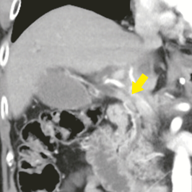 Fig. 1
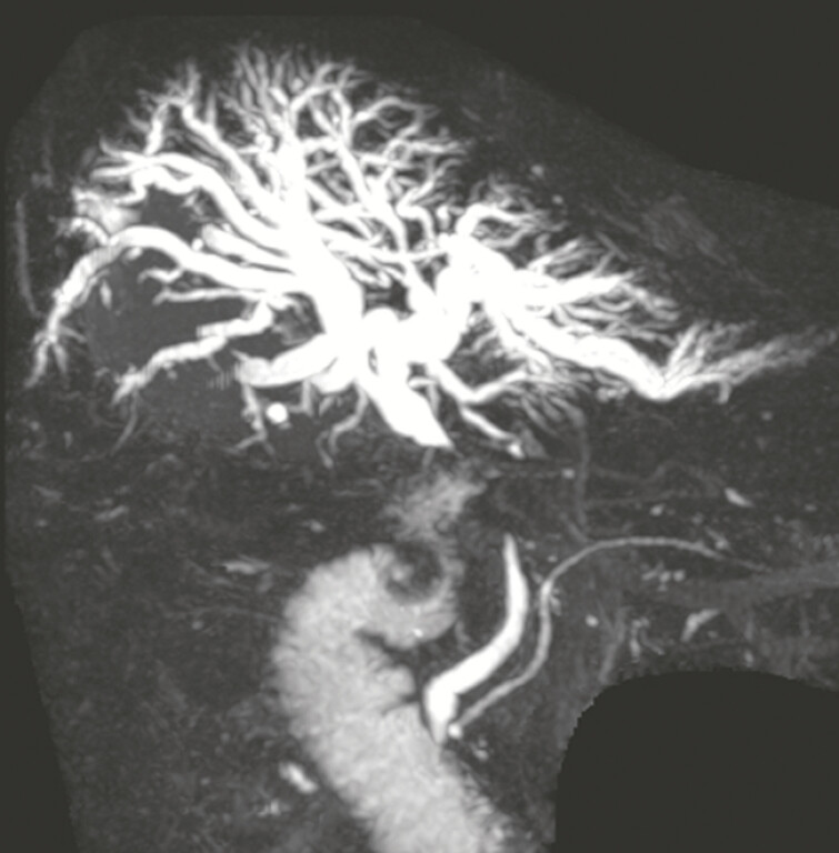 Fig. 2
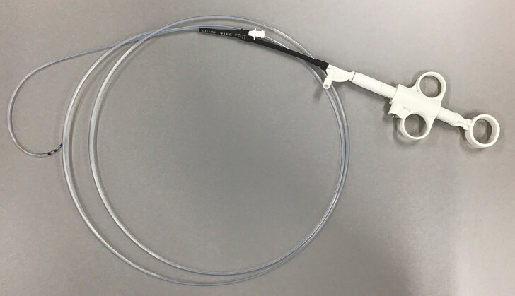 Fig. 3
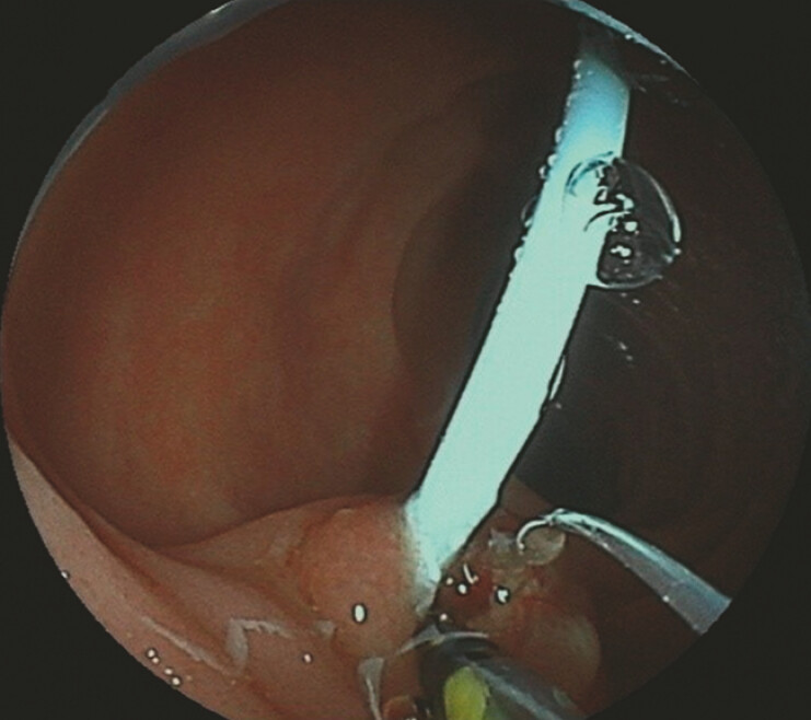 Fig. 4
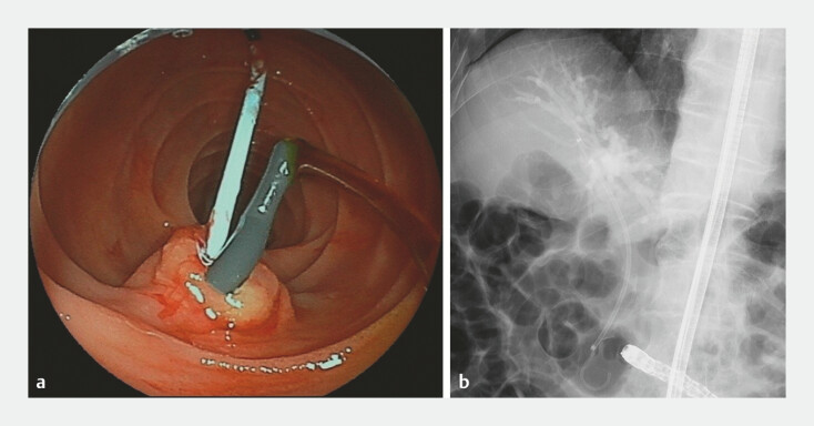 Fig. 5
- —Japan Society for the Promotion of Science10.13039/501100001691
Peer Reviews
No public reviews on file for this paper yet. If you reviewed it on a platform where reviews are public (OpenReview, ICLR, NeurIPS, ICML), you can paste yours below so the community can read it here.
Videos
No videos yet. Explain this paper in a talk, walkthrough, or lecture? Add one.
Taxonomy
TopicsGastrointestinal Bleeding Diagnosis and Treatment · Esophageal and GI Pathology · Gallbladder and Bile Duct Disorders
Bile duct cannulation via balloon endoscopy-assisted endoscopic retrograde cholangiopancreatography (ERCP) can be challenging, particularly in complex anatomical scenarios 1 2 . This case report emphasizes the clinical application of a novel rotatable sphincterotome in a 75-year-old man who presented to a local clinic with a 1-week history of bilirubinuria. The patient had a history of gastric cancer and had undergone a distal gastrectomy with Roux-en-Y reconstruction 6 years previously. Laboratory tests revealed elevated liver enzymes, prompting a referral to our hospital. Contrast-enhanced computed tomography and magnetic resonance cholangiopancreatography revealed mild common bile duct wall thickening and stricture with upstream biliary dilation ( Fig. 1 , Fig. 2 ), which was eventually diagnosed as recurrent gastric cancer 6 months after ERCP.
Contrast-enhanced computed tomography in a patient with a history of gastric cancer treated with distal gastrectomy and Roux-en-Y reconstruction revealed mild thickening and stricture (arrow) of the common bile duct wall and upstream biliary dilation.
Magnetic resonance cholangiopancreatography revealed a stricture of the common bile duct with upstream biliary dilation.
A double-balloon endoscope was used to access the papilla. However, significant challenges prevented successful bile duct cannulation. Retroflex position, a technique often used to facilitate cannulation 3 , was unsuccessful due to the narrow duodenal lumen. Furthermore, conventional sphincterotomy failed as the instrument could not rotate adequately under balloon-assisted endoscopy, and the curvature of the knife did not align with the bile duct axis. Subsequently, a novel, upgraded sphincterotome (Aimingtome; Asahi Intecc Co., Ltd., Seto, Japan) was used ( Fig. 3 ) 4 . This device features a more rotatable and flexible tip, which enabled guidewire insertion into the duodenal papilla ( Video 1 ). The guidewire was then successfully advanced into the main pancreatic duct, facilitating bile duct cannulation via the pancreatic duct guidewire technique. Endoscopic sphincterotomy was performed using the same sphincterotome ( Fig. 4 ), followed by the placement of a biliary plastic stent ( Fig. 5 ). The patient was discharged 3 days after the procedure. In cases where frontal visualization of the papilla using balloon endoscopy-assisted ERCP is challenging, the use of a novel rotatable sphincterotome can effectively facilitate bile duct cannulation and subsequent endoscopic sphincterotomy.
Macroscopic overview of the novel sphincterotome. Source: Asahi Intecc, Seto, Japan.
An upgraded rotatable sphincterotome successfully facilitated bile duct cannulation using balloon endoscopy-assisted endoscopic retrograde cholangiopancreatography. Source for sphincterotome: Asahi Intecc, Seto, Japan.Video 1
Endoscopic sphincterotomy using the novel sphincterotome. Compared to a conventional sphincterotome, it allows 360° rotation and greater backward flexibility.
Biliary and pancreatic stents placed in the common bile duct and main pancreatic duct: a endoscopic view; b radiographic image
Endoscopy_UCTN_Code_TTT_1AR_2AC
The reference list from the paper itself. Each links out to its DOI / PubMed record.
- 1Skinner M Popa D Neumann HERCP with the overtube-assisted enteroscopy technique: a systematic review Endoscopy 20144656057224839188 10.1055/s-0034-1365698 · doi ↗ · pubmed ↗
- 2Martínez-Alcalá García A Aedtner FMönkemüller K Balloon-assisted enteroscopy-ERCP with percutaneous transhepatic rendezvous technique for placement of a self-expanding metal stent Endoscopy 202355 E 930E 93110.1055/a-2119-087537500087 PMC 10374395 · doi ↗ · pubmed ↗
- 3Shimatani M Mitsuyama T Yamashina T Advanced technical tips and recent insights in ERCP using balloon-assisted endoscopy DEN Open 20244 e 30110.1002/deo 2.30138023665 PMC 10644950 · doi ↗ · pubmed ↗
- 4Hori Y Hayashi K Naitoh I Feasibility of newly designed rotatable sphincterotome for endoscopic sphincterotomy (with video)Endosc Int Open 202412 E 1374 E 137839610948 10.1055/a-2422-2425 PMC 11604308 · doi ↗ · pubmed ↗