Multimodal Imaging Characteristics in Unilateral Occlusive Macular Telangiectasia with Atypical X-Shaped Lesion
Abdullah Ağın, Ilknur Turk, Burcu Yakut

TL;DR
This paper describes a rare case of a unique X-shaped lesion in a type of retinal disease called MacTel, using multiple imaging techniques to better understand its characteristics.
Contribution
The paper introduces a novel X-shaped lesion pattern in Type 3a MacTel, expanding the known imaging spectrum of the disease.
Findings
Multimodal imaging revealed an X-shaped hypopigmented lesion with central pigmentation and ischemic features.
OCTA confirmed right-angled venules and localized ischemia in the deep capillary plexus.
The case supports the diagnosis of Type 3a MacTel with distinct vascular and structural abnormalities.
Abstract
Macular Telangiectasia (MacTel) is a rare retinal vascular disorder, with Type 3a MacTel being a distinct form characterized by retinal ischemia with the classical findings of MacTel, such as juxtafoveal telangiectasis, right-angled venules, and deep capillary plexus involvement without central nervous system findings. This case presents a novel X-shaped lesion pattern and ischemic features, expanding the known imaging spectrum of MacTel. A 53-year-old male with diabetes and a history of aripiprazole use presented with persistent blurred vision, a black curtain sensation, and metamorphopsia in the right eye. Visual acuity was 0.8 in the right eye and 1.0 in the left. A multimodal imaging approach, including fundus photography, fundus autofluorescence (FAF), fluorescein angiography (FFA), optical coherence tomography (OCT), and optical coherence tomography angiography (OCTA), was used to…
Genes, proteins, chemicals, diseases, species, mutations and cell lines named across the full text — each resolved to its canonical identifier and authoritative record.
Click any figure to enlarge with its caption.
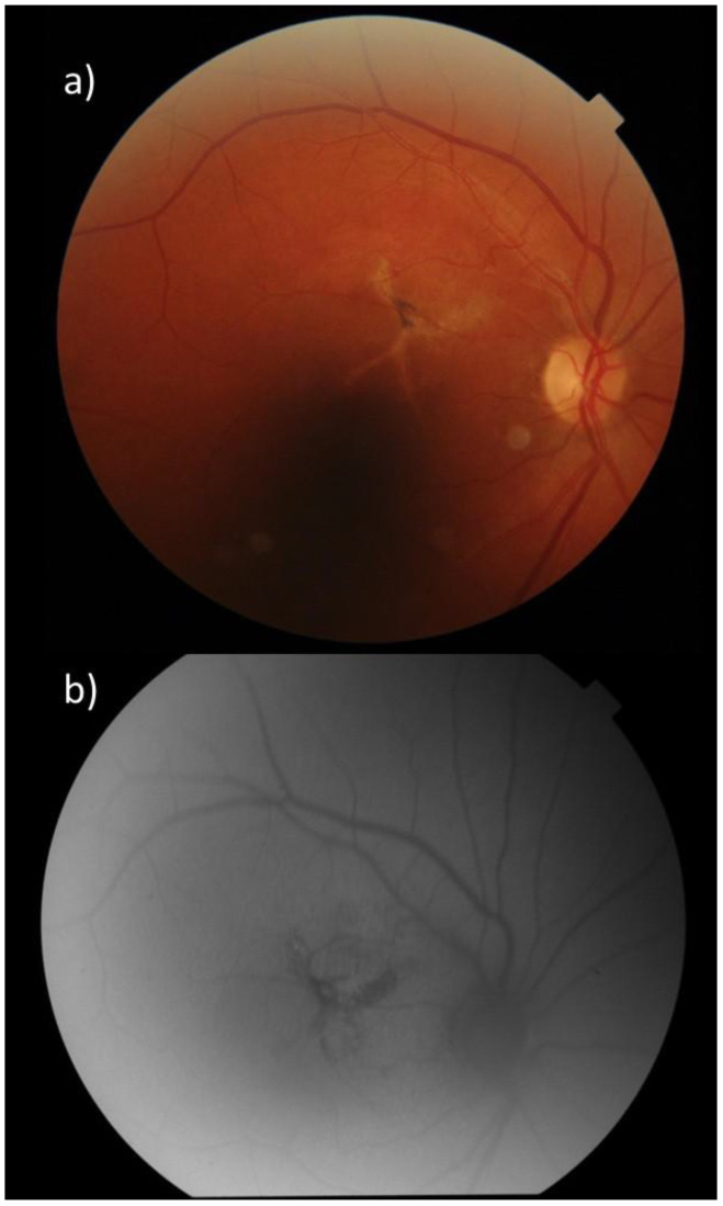 Figure 1
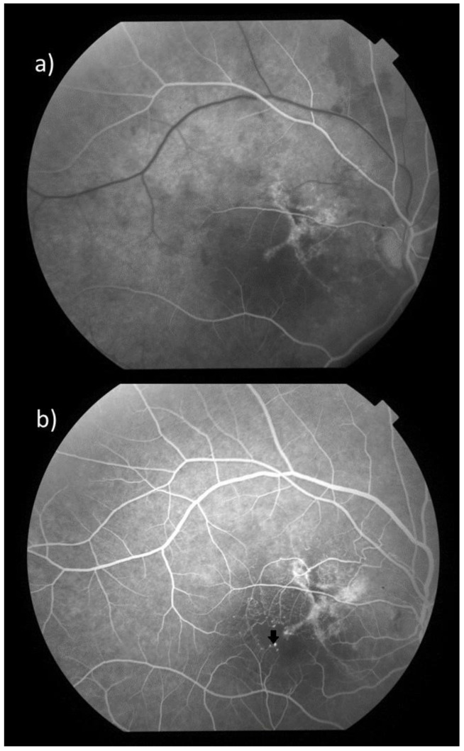 Figure 2
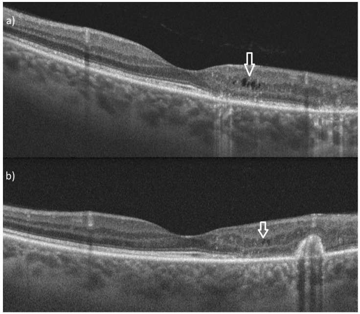 Figure 3
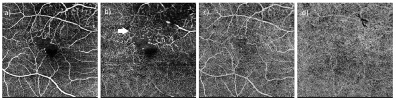 Figure 4
Peer Reviews
No public reviews on file for this paper yet. If you reviewed it on a platform where reviews are public (OpenReview, ICLR, NeurIPS, ICML), you can paste yours below so the community can read it here.
Videos
No videos yet. Explain this paper in a talk, walkthrough, or lecture? Add one.
Taxonomy
TopicsRetinal Diseases and Treatments · Retinal and Optic Conditions · Retinal Imaging and Analysis
The reference list from the paper itself. Each links out to its DOI / PubMed record.
- 1Gass J.D. Blodi B.A. Idiopathic juxtafoveolar retinal telangiectasis. Update of classification and follow-up study Ophthalmology 19931001536154610.1016/S 0161-6420(93)31447-88414413 · doi ↗ · pubmed ↗
- 2Yannuzzi L.A. Bardal A.M. Freund K.B. Chen K.J. Eandi C.M. Blodi B. Idiopathic macular telangiectasia Arch. Ophthalmol.200612445046010.1001/archopht.124.4.45016606869 · doi ↗ · pubmed ↗