Exceptional localization of intramuscular hemangioma: Insights from a 66-year-old case
Joud Boutaleb, Basma Beqqali, Sarah Loubaris, Manal El Beyeg, Ouijdane Zamani, Znati Kaoutar, Rachida Saouab, Jamal El Fenni

TL;DR
A rare case of a benign muscle tumor in a 66-year-old patient is reported, showing how it developed over ten years in the calf muscle.
Contribution
The novelty lies in the detailed clinical and diagnostic description of an intramuscular hemangioma with a long progression timeline.
Findings
The tumor originated from the flexor hallucis longus muscle and caused progressive swelling and pain.
MRI was used to accurately characterize the lesion, supporting the diagnosis of intramuscular hemangioma.
Surgical resection is highlighted as the recommended treatment for such cases.
Abstract
Intramuscular hemangiomas are rare benign vascular tumors, accounting for less than 1% of all hemangiomas. They often present with subtle symptoms that gradually worsen over time. Magnetic resonance imaging (MRI) is the gold standard for diagnosis, providing detailed lesion characterization. Surgical resection remains the preferred treatment. We report the case of a 66-year-old patient with a painful swelling in the calf that had progressively increased in size over ten years, originating from the flexor hallucis longus muscle.
Genes, proteins, chemicals, diseases, species, mutations and cell lines named across the full text — each resolved to its canonical identifier and authoritative record.
Click any figure to enlarge with its caption.
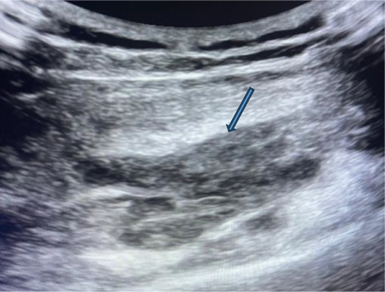 Figure 1
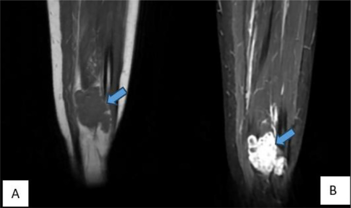 Figure 2
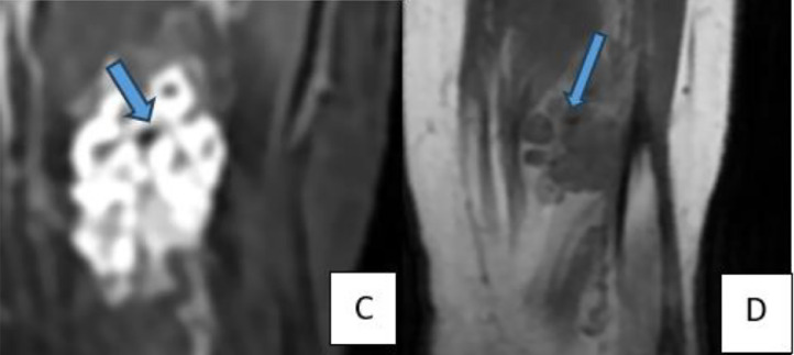 Figure 3
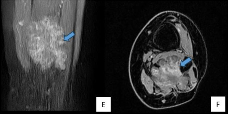 Figure 4
 Figure 5
Peer Reviews
No public reviews on file for this paper yet. If you reviewed it on a platform where reviews are public (OpenReview, ICLR, NeurIPS, ICML), you can paste yours below so the community can read it here.
Videos
No videos yet. Explain this paper in a talk, walkthrough, or lecture? Add one.
Taxonomy
TopicsVascular Malformations and Hemangiomas · Vascular Malformations Diagnosis and Treatment · Ear and Head Tumors