Impact of double-bolus tracking to individualize scan timing of the portal venous phase in preoperative computed tomography colonography angiography for right-sided colon cancer
Yoshiya Ohashi, Masaaki Miyo, Koichi Okuya, Emi Akizuki, Atsushi Hamabe, Ai Noda, Masayuki Ishii, Ryo Miura, Momoko Ichihara, Maho Toyota, Kohei Okamoto, Shun Hayasaka, Takeo Tanaka, Hiroyuki Takashima, Kohei Harada, Keishi Ogura, Ichiro Takemasa, Tsutomu Kumamoto

TL;DR
A new method called double-bolus tracking improves the timing of scans in CT colonography for better imaging of blood vessels in right-sided colon cancer patients.
Contribution
The double-bolus tracking method individualizes scan timing, enhancing contrast and image quality in portal venous phase imaging.
Findings
CT values in the DBT group were significantly higher than in the fixed-delay group for multiple veins.
Visual assessment scores for the gastrocolic trunk were significantly better in the DBT group.
The DBT method improves contrast effect and image quality in portal venous systems.
Abstract
In computed tomography colonography angiography (CTC-A), used for preoperative screening of right-sided colon cancer, the timing of venous phase imaging is conventionally determined by a fixed-delay time; however, the contrast effect may be insufficient because of individual differences in blood flow status. Therefore, we developed the double-bolus tracking (DBT) method to solve this issue. We compared the contrast effect and image quality of the portal venous systems between two methods of the conventional fixed-delay and DBT which utilizes low-dose monitoring to individualize venous scan timings. Data from 30 consecutive patients who underwent CTC-A for right-sided colon cancer using the DBT method were prospectively collected and compared with that from 30 consecutive patients who underwent the conventional fixed-delay method between August 2018 and July 2022. CT values of the…
Genes, proteins, chemicals, diseases, species, mutations and cell lines named across the full text — each resolved to its canonical identifier and authoritative record.
Click any figure to enlarge with its caption.
 Figure 1
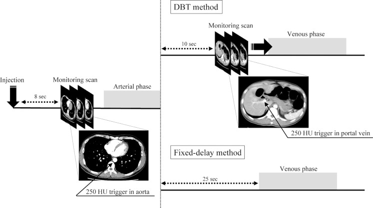 Figure 2
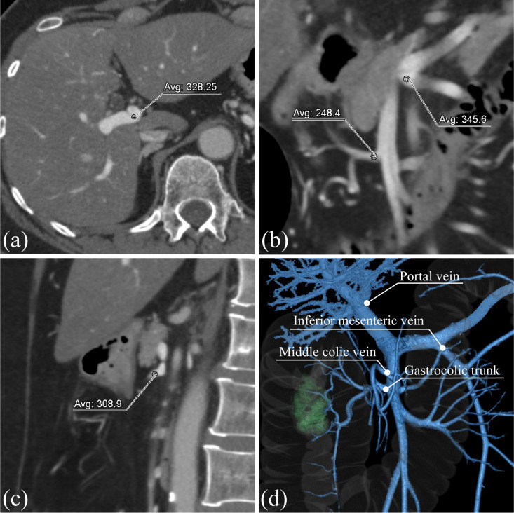 Figure 3
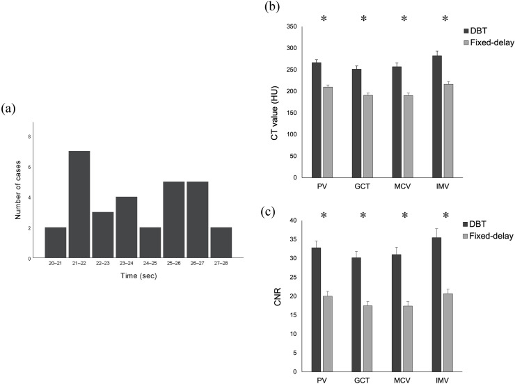 Figure 4
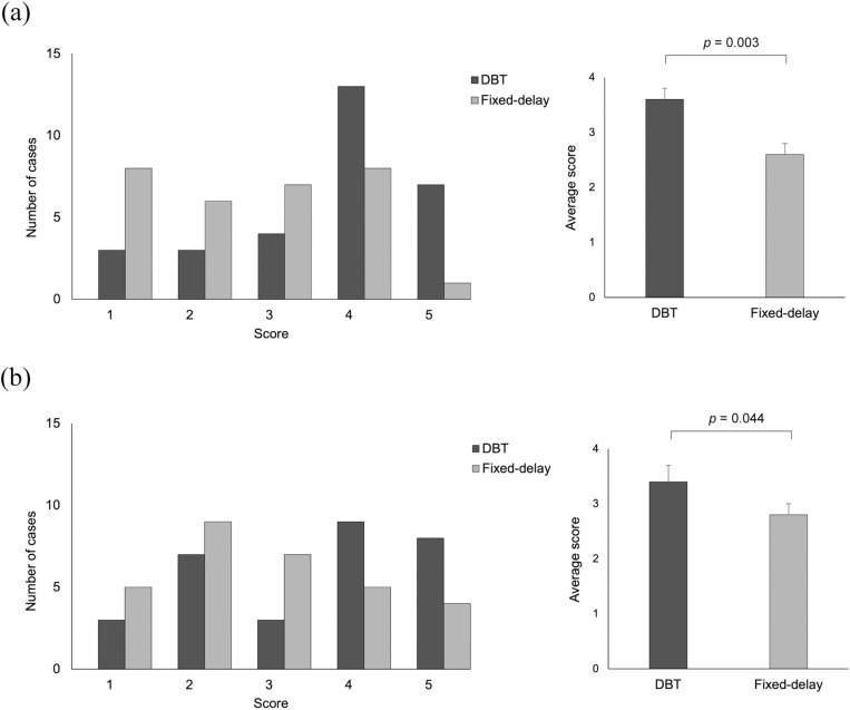 Figure 5
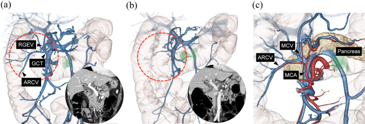 Figure 6
 Figure 7
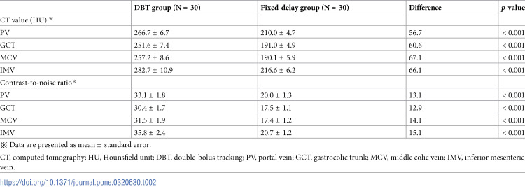 Figure 8
Peer Reviews
No public reviews on file for this paper yet. If you reviewed it on a platform where reviews are public (OpenReview, ICLR, NeurIPS, ICML), you can paste yours below so the community can read it here.
Videos
No videos yet. Explain this paper in a talk, walkthrough, or lecture? Add one.
Taxonomy
TopicsColorectal Cancer Surgical Treatments · Colorectal Cancer Screening and Detection · Advanced X-ray and CT Imaging