High-resolution multi-modal imaging of sub-cellular structures with low numerical aperture objective
Somaiyeh Khoubafarin, Peuli Nath, Saloni Malla, Durgesh Desai, William D Gorgas, Amit K Tiwari, Aniruddha Ray

TL;DR
This paper introduces a cost-effective method to achieve high-resolution imaging of subcellular structures using low numerical aperture objectives, enabling affordable and portable multi-modal microscopy.
Contribution
The novel use of a 2D microlens substrate to enhance resolution and capture evanescent waves with low N.A. objectives.
Findings
Sub-diffraction-limited resolution (<400 nm) was achieved using a 0.25 N.A. objective with a microlens substrate.
The method enables simultaneous scattering, phase, and fluorescence imaging of breast cancer cells and nanoparticle uptake.
The approach improves light capture efficiency and resolution by collecting evanescent waves.
Abstract
Imaging of subcellular structures, which underpins many of the advances in biological and medical sciences, requires microscopes with high numerical aperture (N.A.) objectives which are costly, complex, requires oil immersion and have very limited field-of-view, typically covering a handful of cells. Here, we leverage a low N.A. objective to simultaneously capture scattering, phase, and fluorescence images of subcellular structures in breast cancer cells (BT-20) and observe nanoparticle uptake, with sub-diffraction-limited resolution (<400 nm with a 0.25 N.A. objective) utilizing a 2-dimensional (2-D) microlens substrate. High resolution labeled and label-free images of subcellular components is made possible by implementing a specific configuration, wherein the sample is placed in close proximity to the microlens substrate, which results in efficient collection of the rapidly decaying…
Genes, proteins, chemicals, diseases, species, mutations and cell lines named across the full text — each resolved to its canonical identifier and authoritative record.
Click any figure to enlarge with its caption.
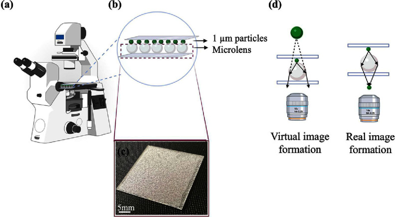 Figure 1
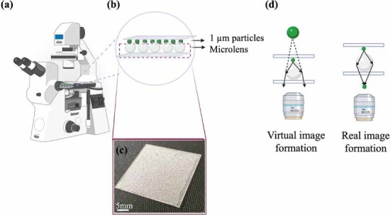 Figure 2
 Figure 3
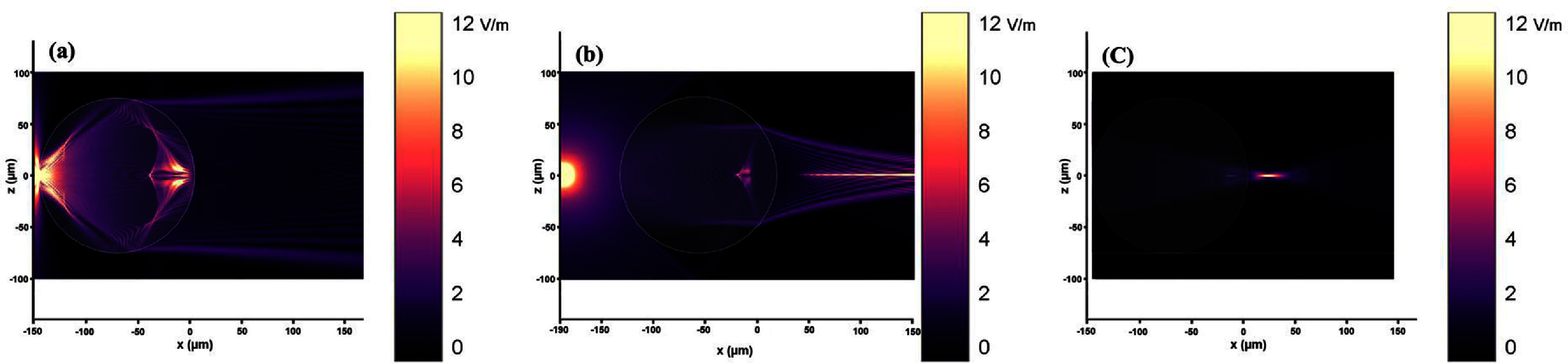 Figure 4
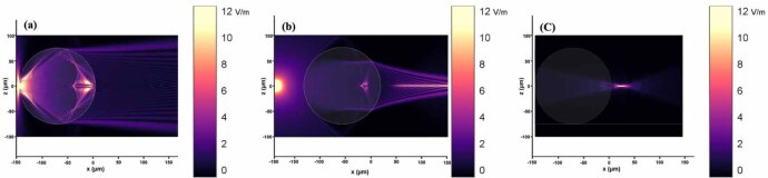 Figure 5
 Figure 6
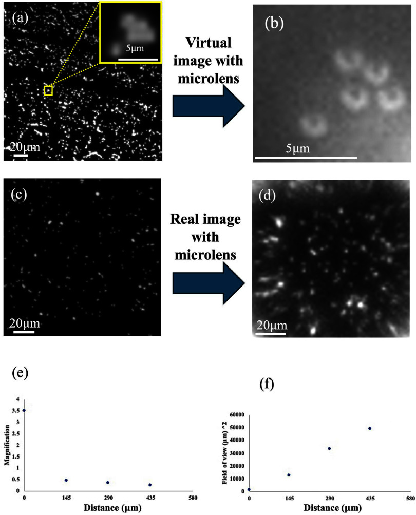 Figure 7
 Figure 8
 Figure 9
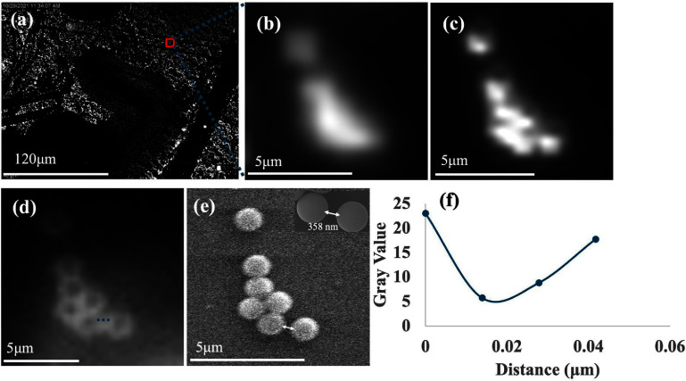 Figure 10
 Figure 11
 Figure 12
 Figure 13
 Figure 14
 Figure 15
 Figure 16
 Figure 17
 Figure 18
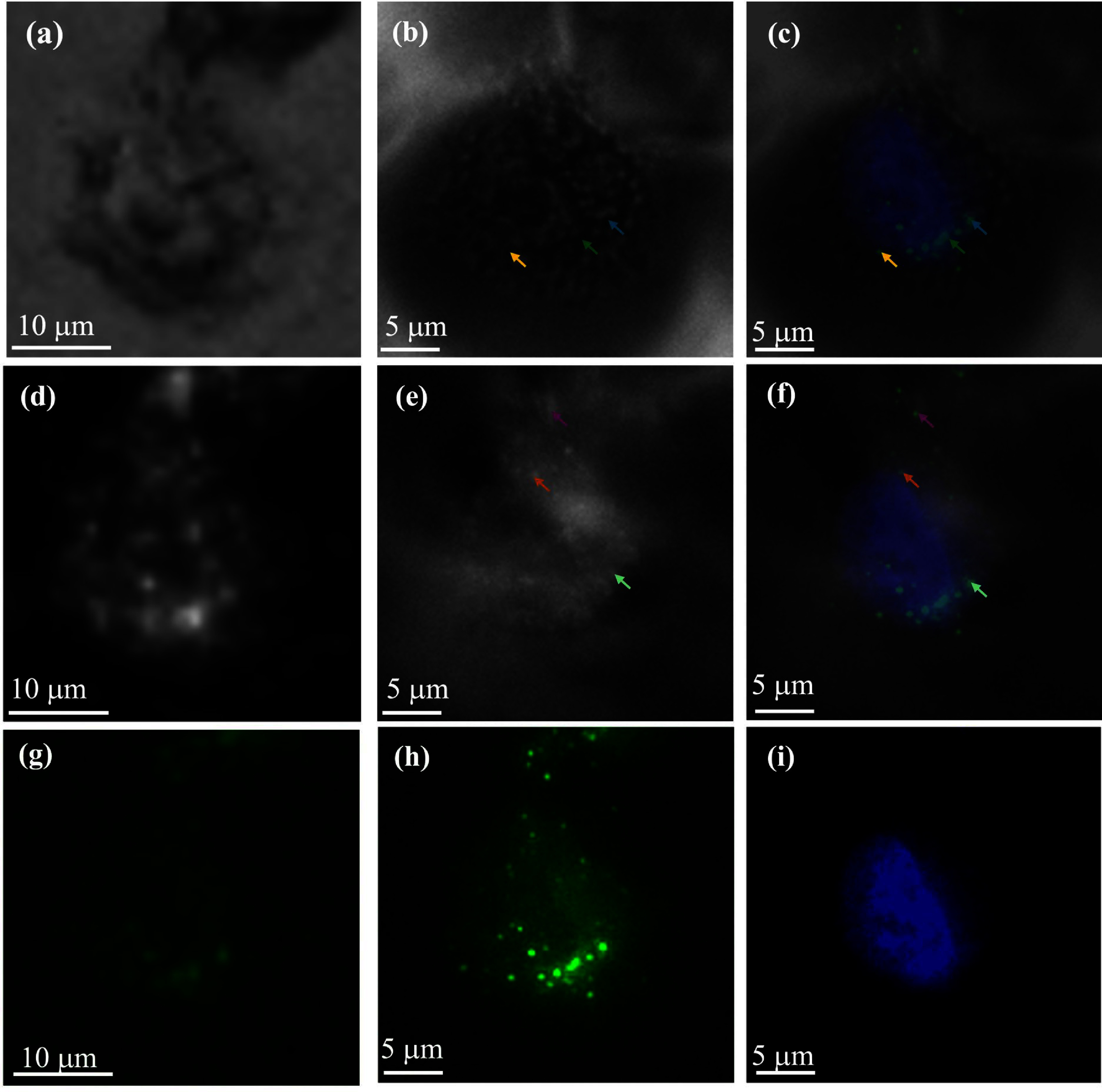 Figure 19
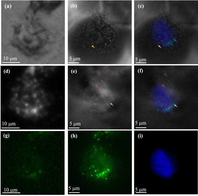 Figure 20
 Figure 21
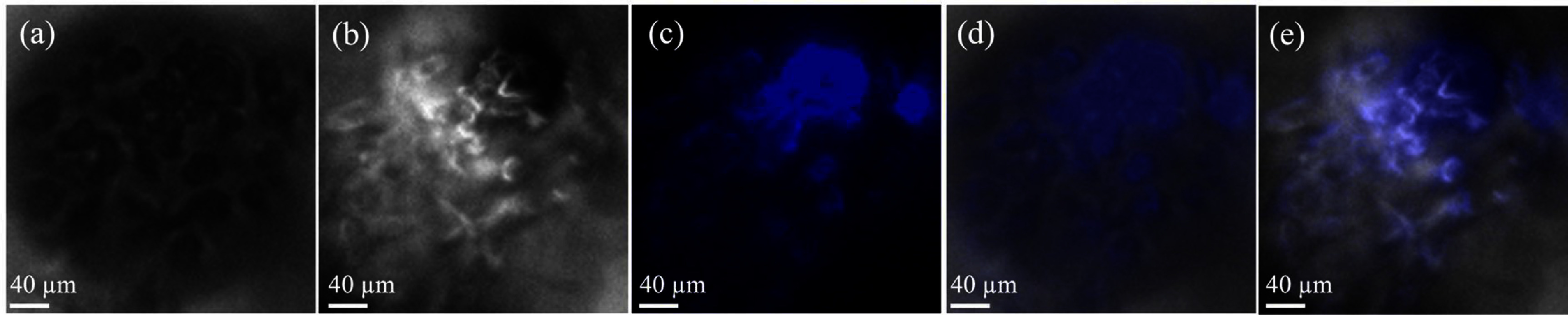 Figure 22
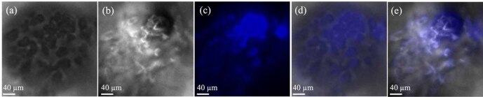 Figure 23
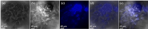 Figure 24
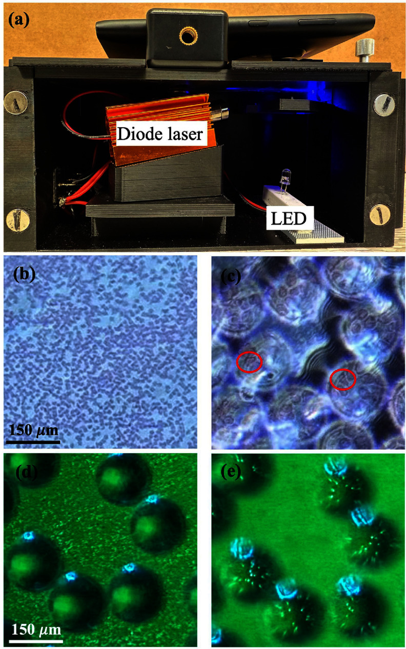 Figure 25
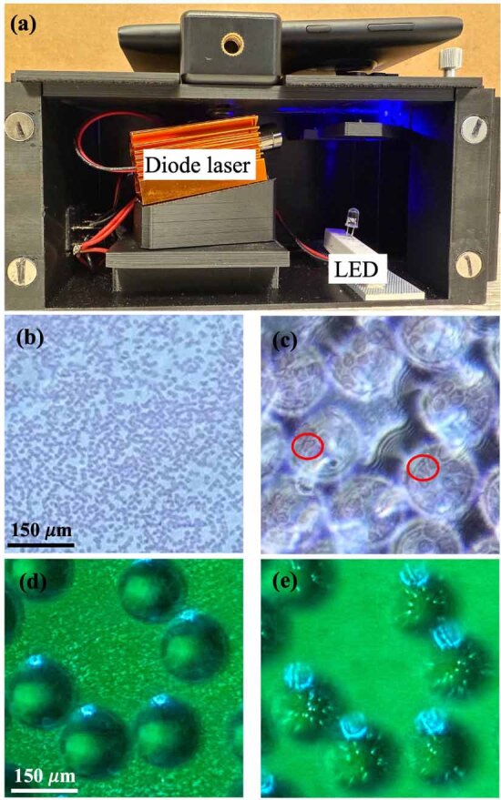 Figure 26
 Figure 27
Peer Reviews
No public reviews on file for this paper yet. If you reviewed it on a platform where reviews are public (OpenReview, ICLR, NeurIPS, ICML), you can paste yours below so the community can read it here.
Videos
No videos yet. Explain this paper in a talk, walkthrough, or lecture? Add one.
Taxonomy
TopicsAdvanced Fluorescence Microscopy Techniques · Photoacoustic and Ultrasonic Imaging · Optical Coherence Tomography Applications