A rare case of MRI-induced thermal burn: Clinical implications and safety awareness
Amer Abu-Shanab, Hamzeh Nasr, Ahmed Mohd, Mahmoud Shaweish, Ahmad Abdulraheem, Doantrang Du

TL;DR
An elderly diabetic woman suffered a rare MRI-induced thermal burn, emphasizing the need for safety protocols and awareness of delayed symptoms.
Contribution
Highlights a rare case of MRI-induced thermal burn and its delayed diagnosis in a diabetic patient.
Findings
A 76-year-old diabetic woman developed a stage III thermal burn after an MRI scan.
The burn was initially misdiagnosed as cellulitis but later identified as MRI-related.
Proper safety measures and patient education are crucial to prevent such injuries.
Abstract
Magnetic Resonance Imaging (MRI) is a widely used, noninvasive diagnostic tool in clinical practice, with millions of scans performed annually. Despite its safety profile, MRI is associated with several potential side effects, including rare but serious complications such as thermal burns. These burns typically result from the interaction of radiofrequency (RF) energy with conductive materials in contact with the skin, or from improper positioning during the scan. This case report details a 76-year-old female who developed a thermal burn on her left elbow after undergoing an MRI of her right shoulder. The patient initially experienced localized heat, redness, and mild tenderness at the site, which later developed into an open wound, leading to a diagnosis of cellulitis. However, further evaluation revealed the wound to be a stage III burn, likely resulting from RF energy exposure during…
Genes, proteins, chemicals, diseases, species, mutations and cell lines named across the full text — each resolved to its canonical identifier and authoritative record.
Click any figure to enlarge with its caption.
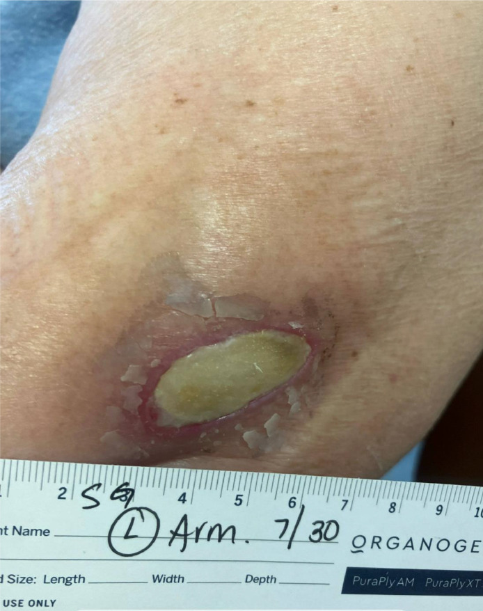 Figure 1
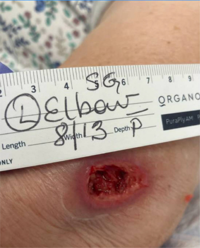 Figure 2
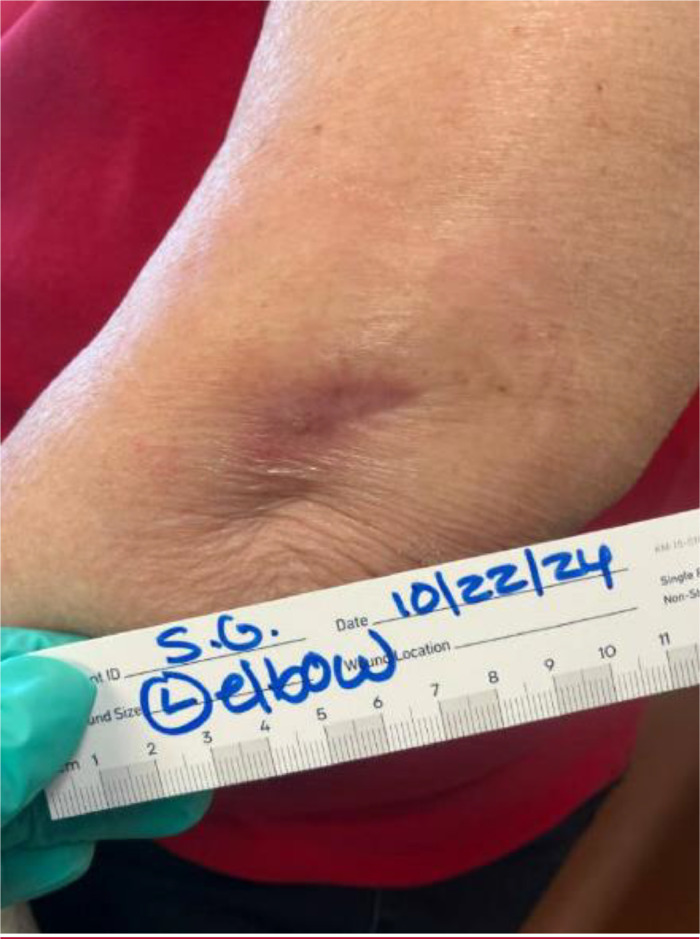 Figure 3
Peer Reviews
No public reviews on file for this paper yet. If you reviewed it on a platform where reviews are public (OpenReview, ICLR, NeurIPS, ICML), you can paste yours below so the community can read it here.
Videos
No videos yet. Explain this paper in a talk, walkthrough, or lecture? Add one.
Taxonomy
TopicsUltrasound and Hyperthermia Applications · Microwave Imaging and Scattering Analysis · Ultrasonics and Acoustic Wave Propagation
Background/Introduction
Magnetic Resonance Imaging (MRI) has emerged as a valuable diagnostic tool in clinical applications since the mid-1980′s. MRI is generally a safe, noninvasive diagnostic technique with nearly 40 million scans carried out annually in the United States [1]. However, like any medical procedure, it is not without potential side effects. Common side effects of MRI include claustrophobia [2], noise-induced discomfort [3], and dizziness or nausea [1]. More serious but rare side effects include allergic reactions to contrast agents and nephrogenic systemic fibrosis in patients with severe kidney dysfunction [4,5].
Thermal burns, although rare, represent a potentially serious complication of MRI [6]. Of MRI adverse effects, 59% that are reported to the Food and Drug Administration (FDA) include thermal injuries [7]. The exact prevalence of MRI-induced burns is difficult to determine due to underreporting [7]. These burns typically result from the interaction between radiofrequency (RF) energy and conductive materials in contact with the patient's skin [8]. This case report presents a rare instance of an MRI-induced skin burn, highlighting the importance of adherence to safety protocols and the need for increased awareness among healthcare professionals and patients about this uncommon but potentially serious complication of MRI procedures.
Case presentation
This case presents a 76-year-old female patient, with a past medical history of diabetes malleus type 2, hypertension, hypothyroidism, and migraine disorder, who presented to the emergency department (ED) for redness and pain at the left elbow. Two months prior to her presentation, the patient fell down from her bed injuring her right shoulder. Since the incident, she had pain in her shoulder for which she had an X-ray which was unraveling. The patient was unable to raise her right arm above 90 degree that was associated with pain with passive range of motion at the right shoulder. The empty can test was positive and rotator cuff tear was suspected for which MRI of the right shoulder was ordered.
One week prior to today's presentation she underwent an MRI for the right shoulder. During the imaging, she felt heat at her left elbow and right after the imaging, she noticed redness and mild tenderness at the same site. She ignored it initially hoping that it will go away. Later, she presented to the ED with an open wound at the elbow with worsening in the redness around it. During examination, an elliptical shaped scab to the left elbow with moderate surrounding erythema, without purulence or discharge was noted. Figures 1, 2, and 3 demonstrate the lesion found on the left elbow of the patient. She denied fever, chills, rigors, and fatigue. She was diagnosed with cellulitis and discharged from the ED on cephalexin, with a referral to the wound care clinic.Fig. 1. Left elbow thermal burn lesion on presentation.Fig 1:Fig. 2. Left elbow thermal burn lesion after debridement and 2 weeks of antibiotics.Fig 2:Fig. 3. Left elbow thermal burn lesion after debridement and 2 months of antibiotics.Fig 3:
She took her medication as prescribed and presented to the wound clinic for follow up. On follow up, she had a nonhealing 4 × 2 cm wound with a thick scab. The lesion was red and swollen but not tender. There was no fluctuance nor discharge. She was diagnosed with stage III burns with thick eschar. The X-ray of the left elbow was normal. The wound was debrided, and she was started on Bactrim and topical gentamicin with close follow ups. Sterile dressing was done on a weekly base by the wound care team. The burn was improving over time and was in a healing phase after 8 weeks of dressing and topical antibiotics.
Discussion
This case presents a 76-year-old female patient experiencing a thermal burn post-MRI imaging. This incident highlights the importance of understanding the mechanisms by which MRI can potentially cause skin burns and the need for stringent safety protocols.
MRI scanners employ powerful radiofrequency (RF) pulses to create images, which causes RF heating. Particularly in areas where a concentrated electromagnetic field is applied, these radiofrequency waves have the potential to heat tissue [8]. It's possible that the patient's left elbow was placed in an area with a strong radiofrequency field in this instance.
Another explanation might be conductive loop formation, where the body part might be positioned in a way that created a conductive loop (e.g., skin-to-skin contact) [6]. A case study published in 2017 by Mandel and her colleagues [6] presented a 48-year-old man who experienced a second degree burn on his thighs bilaterally following an MRI. It was believed that the patient mistakenly pushed his thighs together while inside the scanner, creating a closed loop conduction circuit that led to a thermal burn. The significance of proper posture during MRI is demonstrated by the case.
In addition, the presence of undetectable metallics in clothing could be a possible source for thermal burn. Previous case studies showed that nondetectable metallic material, which is found in popular athletic clothing presented a serious risk for MRI cutaneous burns. The metallic fiber in the clothing is difficult to detect by standard MRI screening methods [9,10]. In this case study [10], an MRI was performed on a 40-year-old woman to evaluate her thigh muscles. Following the MRI, both her thighs showed linear redness and swelling that matched the vertical lines on the jogging clothes she was wearing. The redness eventually turned into blistering eruptions a week later. The jogging trousers' material list mentioned 100% polyester and did not include any mention of metal. They later found that the vertical lines of the jogging pants might have been made of light, thin metal fibers after contacting the company's customer service.
Impaired thermoregulation in elderly, particularly those with diabetes, may have contributed to reduced peripheral sensation and impaired thermoregulation [11]. These factors increased the possibility of thermal injury in our patient. Finally, despite our patient denying that, but the application of a cream or a lotion that contains heavy material might increase the absorption of the RF, thus increase the temperature of the area.
The delayed presentation and initial misdiagnosis as cellulitis underscore the importance of post-MRI patient education and follow-up. Patients should be instructed to report any unusual sensations or skin changes promptly after an MRI procedure.
Conclusion
This case emphasizes the need for increased awareness among healthcare providers about the potential for MRI-induced burns, even in the absence of obvious risk factors. It also highlights the importance of prompt recognition and appropriate management of such injuries to prevent complications and ensure optimal patient outcomes.
Patient consent
The patient provided written informed consent to publish the information of her post-MRI burn and any accompanying images.
The reference list from the paper itself. Each links out to its DOI / PubMed record.
- 1Shah A.Aran S.A review of Magnetic resonance (MR) safety: the essentials to patient safety Cureus 15102023 e 4734510.7759/cureus.4734538021512 PMC 10657250 · doi ↗ · pubmed ↗
- 2Kilborn L.C.LabbéE.E.Magnetic resonance imaging scanning procedures: development of phobic response during scan and at one-month follow-up J Behav Med 134199039140110.1007/BF 008448862246785 · doi ↗ · pubmed ↗
- 3Alibek S.Vogel M.Sun W.Winkler D.Baker C.A.Burke M.Acoustic noise reduction in MRI using silent scan: an initial experience Diagn Interv Radiol 204201436036310.5152/dir.2014.1345824808439 PMC 4463276 · doi ↗ · pubmed ↗
- 4Chalise S.N.Palmer E.Pathak V.Fatal allergic reaction to gadolinium contrast Cureus 1572023 e 4245510.7759/cureus.4245537637616 PMC 10449612 · doi ↗ · pubmed ↗
- 5Lim Y.J.Bang J.Ko Y.Seo H.M.Jung W.Y.Yi J.H.Late onset nephrogenic systemic fibrosis in a patient with stage 3 chronic kidney disease: a case report J Korean Med Sci 35352020 e 29310.3346/jkms.2020.35.e 29332893521 PMC 7476800 · doi ↗ · pubmed ↗
- 6Mandel N.S.Ramdial J.L.Marcus E.N.A second-degree burn after MRI Cleve Clin J Med 845201734834910.3949/ccjm.84a.1516428530901 · doi ↗ · pubmed ↗
- 7Baker C.Nugent B.Grainger D.Hewis J.Malamateniou C.Systematic review of MRI safety literature in relation to radiofrequency thermal injury prevention J Med Radiat Sci 713202444546010.1002/jmrs.80038937923 PMC 11569411 · doi ↗ · pubmed ↗
- 8Tang M.Yamamoto T.Progress in understanding radiofrequency heating and burn injuries for safer MR imaging Magn Reson Med Sci 221202372510.2463/mrms.rev.2021-004735228437 PMC 9849420 · doi ↗ · pubmed ↗