Correction to: The role of EMILIN-1 in the osteo/odontogenic differentiation of dental pulp stem cells
Pingmeng Deng, Jing Huang, Qixuan Zhang, Yuejia Li, Jie Li

Abstract
Genes, proteins, chemicals, diseases, species, mutations and cell lines named across the full text — each resolved to its canonical identifier and authoritative record.
Click any figure to enlarge with its caption.
 Figure 1
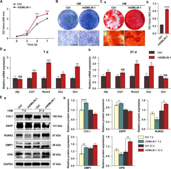 Figure 2
Peer Reviews
No public reviews on file for this paper yet. If you reviewed it on a platform where reviews are public (OpenReview, ICLR, NeurIPS, ICML), you can paste yours below so the community can read it here.
Videos
No videos yet. Explain this paper in a talk, walkthrough, or lecture? Add one.
Taxonomy
TopicsBone and Dental Protein Studies · dental development and anomalies
Correction to: BMC Oral Health (2023) 23:203
10.1186/s12903-023-02905-3
In this article [1], the representative microscopic image of the rhEMILIN-1 treated group in Fig. 6Ca was mistakenly duplicated from the control group image. The incorrect and corrected versions of Fig. 6 are displayed below.
Incorrect Fig. 6.
Fig. 6rhEMILIN-1 coating stimulated osteo/odontogenic differentiation of hDPSCs. A CCK8 assay showed that rhEMILIN-1 treatment significantly increased the proliferation of hDPSCs. B ALP staining (7 days) showed that rhEMILIN-1 decreased ALP activity in hDPSCs. Scale bars = 250 μm. C ARS staining (21 days) showed that rhEMILIN-1 treatment significantly increased the mineralization capacity of hDPSCs (a). Mineralized nodules was quantified (b). Scale bars = 250 μm. D qPCR detected that rhEMILIN-1 treatment increased the relative mRNA expression of osteo/odonto-specific genes at osteo/odontogenesis day 7 (a) and osteo/odontogenesis day 21(b). E Western blot detected that rhEMILIN-1 treatment increased the expression of osteo/odonto-specific proteins in hDPSCs at early and late stages of induction (a). Grayscale analysis of protein bands (b). Values are presented as mean ± standard deviation (SD). *P < 0.05; **P < 0.01; ***P < 0.001; ****P < 0.0001. The blots in Ea were cropped
Correct Fig. 6.
Fig. 6rhEMILIN-1 coating stimulated osteo/odontogenic differentiation of hDPSCs. A CCK8 assay showed that rhEMILIN-1 treatment significantly increased the proliferation of hDPSCs. B ALP staining (7 days) showed that rhEMILIN-1 decreased ALP activity in hDPSCs. Scale bars = 250 μm. C ARS staining (21 days) showed that rhEMILIN-1 treatment significantly increased the mineralization capacity of hDPSCs (a). Mineralized nodules was quantified (b). Scale bars = 250 μm. D qPCR detected that rhEMILIN-1 treatment increased the relative mRNA expression of osteo/odonto-specific genes at osteo/odontogenesis day 7 (a) and osteo/odontogenesis day 21(b). E Western blot detected that rhEMILIN-1 treatment increased the expression of osteo/odonto-specific proteins in hDPSCs at early and late stages of induction (a). Grayscale analysis of protein bands (b). Values are presented as mean ± standard deviation (SD). *P < 0.05; **P < 0.01; ***P < 0.001; ****P < 0.0001. The blots in Ea were cropped