Low lateral inclination angle, high sulcus angle, high trochlear height and patella alta are risk factors for first lateral patellar dislocation and complete MPFL rupture, comparative study
Serhat Akcaalan, Ismail Duran, Abdurrahim Kavaklilar, Fatih Beser, Ceyhun Caglar, Mahmut Ugurlu

TL;DR
This study identifies specific anatomical risk factors for a complete MPFL rupture after a first patellar dislocation, using MRI measurements to predict the likelihood of injury.
Contribution
The study introduces a predictive model using MRI-based anatomical parameters to assess the risk of complete MPFL rupture after first lateral patellar dislocation.
Findings
Low lateral trochlear inclination (LTI) and high sulcus angle (SA) are significant risk factors for complete MPFL rupture.
Patella alta and high trochlear height (TH) also increase the risk of MPFL rupture after first dislocation.
The predictive model based on these factors has a 70.4% accuracy in estimating the risk of MPFL rupture.
Abstract
To identify risk factors for complete medial patello‐femoral ligament (MPFL) rupture after first lateral patellar dislocation (LPD) and to develop a model to predict the risk of rupture. Patients who presented with first LPD between February 2019 and June 2024 and were diagnosed with complete MPFL rupture on magnetic resonance imaging (MRI) were retrospectively reviewed. Patients with normal MRI findings in a 1:1 ratio were selected as the control group by computer‐assisted randomisation.All patients in both groups were asked to perform MRI on, tibial tuberosity–trochlear groove (TT–TG) distance, lateral trochlear inclination (LTI) angle, sulcus angle (SA), medial femoral condyle height (MFCH), lateral femoral condyle height (LFCH), trochlear height (TH), patellotrochlear index (PTI), Koshino–Sugimoto Index (KSI), Caton–Deschamps Index (CDI) and Insall–Salvati Index (ISI) were measured…
Genes, proteins, chemicals, diseases, species, mutations and cell lines named across the full text — each resolved to its canonical identifier and authoritative record.
Click any figure to enlarge with its caption.
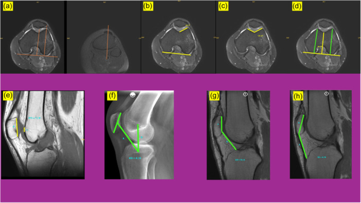 Figure 1
Peer Reviews
No public reviews on file for this paper yet. If you reviewed it on a platform where reviews are public (OpenReview, ICLR, NeurIPS, ICML), you can paste yours below so the community can read it here.
Videos
No videos yet. Explain this paper in a talk, walkthrough, or lecture? Add one.
Taxonomy
TopicsLower Extremity Biomechanics and Pathologies · Foot and Ankle Surgery · Sports injuries and prevention