Evaluation of the Level of Agreement Between Clinical Diagnosis and Two Cephalometric Analyses: Cephalometric Analysis for Orthognathic Surgery (COGS) and Soft Tissue Cephalometric Analysis (STCA)
Ankita Lohia, Siddarth Shetty, Amoli Singh, Shravan Shetty, Ashith M. V.

TL;DR
This study compares two cephalometric analyses, COGS and STCA, to clinical diagnoses in orthognathic surgery, finding COGS more accurate for most parameters except surgical need.
Contribution
The study evaluates diagnostic accuracy of COGS and STCA against clinical diagnoses, revealing their strengths and limitations in orthognathic surgery planning.
Findings
COGS showed better agreement with clinical diagnosis for mandible position and intermaxillary jaw relationship.
STCA performed better than COGS in predicting the need for surgical intervention.
Agreement for other parameters like growth patterns and lip prominence was poor for both analyses.
Abstract
Introduction: Hard tissue analysis, such as cephalometric analysis for orthognathic surgery (COGS), defines the nature of existing skeletal discrepancies but is incomplete in providing information concerning the facial form and proportions of the patient. The soft tissue cephalometric analysis (STCA) accounts for the soft tissue drape, which, however, is subject to significant individual, gender, and age variation. Aims and Objectives: The purpose of the study was to evaluate the conformance of the diagnostic inferences derived from two cephalometric analyses, COGS and STCA, to the clinical diagnosis of experienced clinicians. Material and Methods: Lateral cephalograms of 120 patients were traced for parameters previously diagnosed by an oral surgeon and an orthodontist. Corresponding variables were taken from two analyses, COGS and STCA, defining the (1) position of the maxilla, (2)…
Genes, proteins, chemicals, diseases, species, mutations and cell lines named across the full text — each resolved to its canonical identifier and authoritative record.
Click any figure to enlarge with its caption.
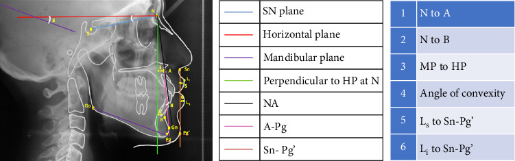 Figure 1
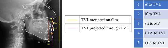 Figure 2
 Figure 3
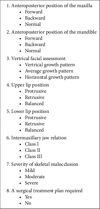 Figure 4
Peer Reviews
No public reviews on file for this paper yet. If you reviewed it on a platform where reviews are public (OpenReview, ICLR, NeurIPS, ICML), you can paste yours below so the community can read it here.
Videos
No videos yet. Explain this paper in a talk, walkthrough, or lecture? Add one.
Taxonomy
TopicsOrthodontics and Dentofacial Orthopedics · Dental Radiography and Imaging · Temporomandibular Joint Disorders