Unveiling CNS cell morphology with deep learning: A gateway to anti-inflammatory compound screening
Hyunseok Bahng, Jung‑Pyo Oh, Sungjin Lee, Jaehong Yu, Jongju Bae, Eun Jung Kim, Sang‑Hun Bae, Ji‑Hyun Lee, Ghulam Md Ashraf, Carla Pegoraro, Carla Pegoraro

TL;DR
This paper introduces a deep learning method to study brain cell shapes, helping identify anti-inflammatory drugs for neurological diseases.
Contribution
The novel contribution is a deep learning approach for analyzing CNS cell morphology to screen anti-inflammatory compounds.
Findings
DL-based analysis of neuronal and microglial cell morphology was successfully applied in pathological conditions.
The method enables efficient screening of therapeutic compounds for neuroinflammation.
The approach overcomes challenges like batch effects and limited labeled data in CNS cell image analysis.
Abstract
Deciphering the complex relationships between cellular morphology and phenotypic manifestations is crucial for understanding cell behavior, particularly in the context of neuropathological states. Despite its importance, the application of advanced image analysis methodologies to central nervous system (CNS) cells, including neuronal and glial cells, has been limited. Furthermore, cutting-edge techniques in the field of cell image analysis, such as deep learning (DL), still face challenges, including the requirement for large amounts of labeled data, difficulty in detecting subtle cellular changes, and the presence of batch effects. Our study addresses these shortcomings in the context of neuroinflammation. Using our in-house data and a DL-based approach, we have effectively analyzed the morphological phenotypes of neuronal and microglial cells, both in pathological conditions and…
Genes, proteins, chemicals, diseases, species, mutations and cell lines named across the full text — each resolved to its canonical identifier and authoritative record.
Click any figure to enlarge with its caption.
 Figure 1
 Figure 2
 Figure 3
 Figure 4
 Figure 5
 Figure 6
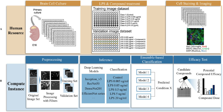 Figure 7
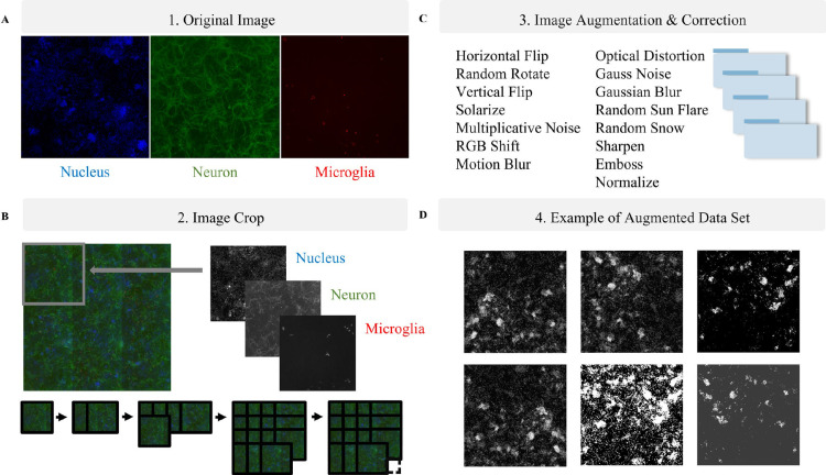 Figure 8
 Figure 9
 Figure 10
 Figure 11
 Figure 12
Peer Reviews
No public reviews on file for this paper yet. If you reviewed it on a platform where reviews are public (OpenReview, ICLR, NeurIPS, ICML), you can paste yours below so the community can read it here.
Videos
No videos yet. Explain this paper in a talk, walkthrough, or lecture? Add one.
Taxonomy
TopicsCell Image Analysis Techniques · Image Processing Techniques and Applications · Digital Imaging for Blood Diseases