Computed tomography–guided percutaneous biopsy in diagnosis of suspected metastatic renal cell carcinoma: which location is the most suitable?
Petr Hoffmann, Michal Balik, Martina Hoffmannova, Jindrich Kopecky, Pavel Ryska, Jana Draganovicova, Petr Dvorak

TL;DR
This study examines the best biopsy location for diagnosing metastatic kidney cancer, finding that biopsies outside the kidney have fewer complications without affecting diagnostic accuracy.
Contribution
The study identifies that non-renal biopsy locations offer lower complication rates without compromising diagnostic accuracy in metastatic renal cell carcinoma.
Findings
Biopsies outside the kidney had a lower complication rate compared to kidney biopsies.
There was no significant difference in diagnostic accuracy between kidney and non-kidney biopsy locations.
Most biopsy results were true-positive, with a small percentage requiring rebiopsy due to false-negative results.
Abstract
Systemic targeted therapy options are commonly used in patients with metastatic renal cell carcinoma (mRCC). Histological verification is crucial for treatment of mRCC. Our aim was to evaluate an optimal location for percutaneous computed tomography‑guided biopsy in a diagnosis of suspected mRCC. A total of 138 percutaneous biopsies for tumors ranging from 21 to 133 mm in diameter (median, 72 mm) were carried out in 134 patients with suspected mRCC over a 5‑year period. The biopsy location was variable, with kidney biopsy performed in 77 cases (55.8%), and other localizations (retroperitoneum, peritoneal cavity, liver, pelvis, pleural space, lung, mediastinum, chest or abdominal wall, and pancreas) in 61 cases (44.2%). As many as 288 biopsies (97.1%), yielded truepositive results, and 4 procedures (2.9%) yielded histologically falsenegative results that required confirmation through…
Genes, proteins, chemicals, diseases, species, mutations and cell lines named across the full text — each resolved to its canonical identifier and authoritative record.
Click any figure to enlarge with its caption.
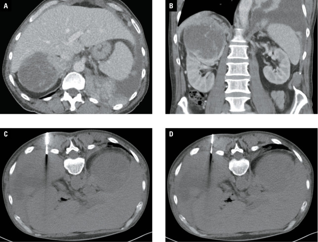 Figure 1
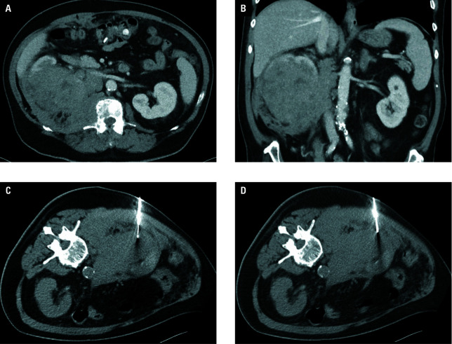 Figure 2
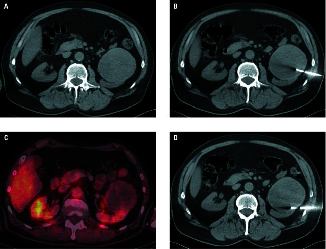 Figure 3
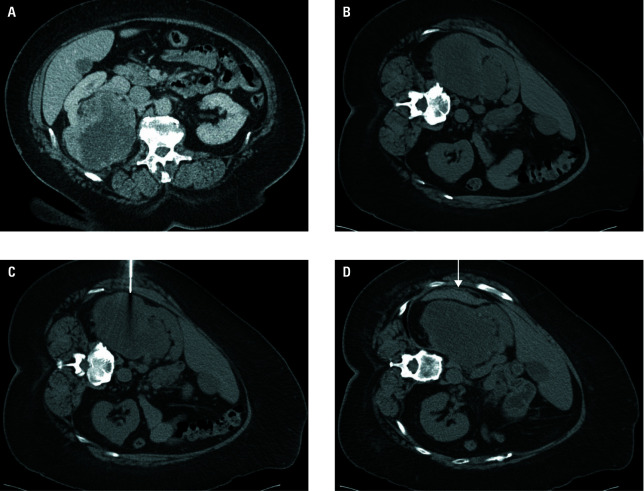 Figure 4
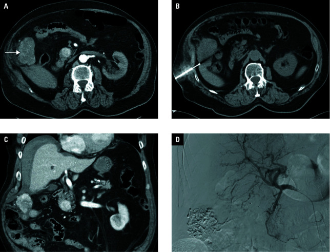 Figure 5
Peer Reviews
No public reviews on file for this paper yet. If you reviewed it on a platform where reviews are public (OpenReview, ICLR, NeurIPS, ICML), you can paste yours below so the community can read it here.
Videos
No videos yet. Explain this paper in a talk, walkthrough, or lecture? Add one.
Taxonomy
TopicsRenal cell carcinoma treatment · Renal and related cancers · Renal and Vascular Pathologies