Exploring Curriculum Considerations to Prepare Future Radiographers for an AI-Assisted Health Care Environment: Protocol for Scoping Review
Chamandra Kammies, Elize Archer, Penelope Engel-Hills, Mariette Volschenk

TL;DR
This study aims to identify how radiography education should change to prepare future radiographers for working with AI in healthcare.
Contribution
The novelty lies in systematically mapping the evidence for curriculum adjustments in radiography education due to AI integration.
Findings
A scoping review is being conducted to explore radiography curriculum changes for AI-assisted healthcare.
Initial database searches yielded 2224 records, with title and abstract screening underway.
The review will provide insights into educational needs for radiographers in an AI-driven healthcare environment.
Abstract
The use of artificial intelligence (AI) technologies in radiography practice is increasing. As this advanced technology becomes more embedded in radiography systems and clinical practice, the role of radiographers will evolve. In the context of these anticipated changes, it may be reasonable to expect modifications to the competencies and educational requirements of current and future practitioners to ensure successful AI adoption. The aim of this scoping review is to explore and synthesize the literature on the adjustments needed in the radiography curriculum to prepare radiography students for the demands of AI-assisted health care environments. Using the Joanna Briggs Institute methodology, an initial search was run in Scopus to determine whether the search strategy that was developed with a library specialist would capture the relevant literature by screening the title and…
Genes, proteins, chemicals, diseases, species, mutations and cell lines named across the full text — each resolved to its canonical identifier and authoritative record.
Click any figure to enlarge with its caption.
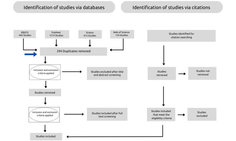 Figure 1
| PCCa Framework | Include | Exclude |
| Population |
All 4 radiography categories Radiography educators Radiography students | All other health professions’ disciplines |
| Concept |
Studies that discuss AIb or AI-based tools in the context of radiography | Studies using technology that does not incorporate AI-based tools |
| Context |
Academic and clinical radiography education Undergraduate radiography education Postgraduate radiography education Continuing professional development | Other health professions’ education programs |
| Study characteristics |
The review will consider all types of study designs. Sources printed in all languages Full-text articles | Full-text not available |
| Electronic resources | Sources, n |
| EBSCO (Academic Search Premier, CINAHL, ERIC) | 362 |
| PubMed | 1214 |
| Scopus | 267 |
| Web of Science | 112 |
| Gray literature | 269 |
| Results | 2224 |
Peer Reviews
No public reviews on file for this paper yet. If you reviewed it on a platform where reviews are public (OpenReview, ICLR, NeurIPS, ICML), you can paste yours below so the community can read it here.
Videos
No videos yet. Explain this paper in a talk, walkthrough, or lecture? Add one.
Taxonomy
TopicsArtificial Intelligence in Healthcare and Education · Radiomics and Machine Learning in Medical Imaging · AI in cancer detection
Introduction
Radiography is arguably one of the most technologically advanced health care disciplines, encompassing various technologies that continuously evolve with advancements in computing power and human knowledge [1,2]. Although the use of technology has always been extensive in the profession, recent technological advancements have focused on incorporating complex machine learning algorithms, which have led to a change in clinical protocols that affect patient outcomes and radiography practice [3-5]. Artificial intelligence (AI) technologies play an increasingly important role in clinical radiography with applications such as scheduling patients, image interpretation and reporting, vetting of examinations, patient positioning, image generation and reconstruction, radiation therapy dosimetry, and image postprocessing [4,6-8]. As the number of AI applications increases in clinical practice [9], it is postulated that the practitioner role may change and expand, and these new roles will require modifications to the competencies and educational requirements of current and future practitioners [4].
The increasing integration of AI technologies in radiography practice raises significant questions about the influences of AI technology on radiography practice and the subsequent ways in which radiography education may be required to change. Recent reviews have explored AI’s current and potential applications in radiography practice [5] and examined the AI educational programs available to radiography staff globally [1]. Furthermore, research undertaken in the United Kingdom, Africa, and the Middle East exploring AI topics linked to education in radiography has focused on equipping practitioners for successful AI adoption [10] as well as the perceptions and attitudes of students and practitioners [11-16]. These conversations illustrate that there has been increasing exploration of various facets of AI in radiography, indicating that the emergence of AI technologies will continue to shape the future of radiography and radiography education.
Opinion statements and discussion pieces argue for the inclusion of AI education in undergraduate radiography curricula and appropriate education for current practitioners [3,4,17]. In addition, recommendations were made that higher education institutions must ensure that radiography curricula provide educational opportunities for patient-centered care in relation to AI integration to ensure competent and safe practice [2,3,6]. With increasing automation in clinical tasks, patient-centered care will gain more importance, highlighting the need for academic curricula to prioritize patient-centered care [2,4]. The evolving landscape of radiography, marked by the increasing integration of AI, raises important questions about how to best prepare future radiographers. Therefore, mapping the existing literature on the way that radiography education may need to change because of the integration of AI technologies in clinical practice is needed to grasp the current state of knowledge on the topic for future educational considerations. The aim of this scoping review is to explore and synthesize the literature on the adjustments needed in the radiography curriculum to prepare radiography students for the demands of AI-assisted health care environments.
Methods
Overview
The review will follow the Joanna Briggs Institute methodology for scoping reviews because it uses a rigorous and logical approach to scoping reviews [18,19]. The methodological guideline delineates 6 steps to map the extent of the literature on the research topic. These include (1) defining the research question, (2) developing the inclusion and exclusion criteria, (3) describing the search strategy, (4) searching the literature, (5) data extraction, and (6) analyzing the evidence and presenting the results [19]. The scoping review protocol is registered on the Open Science Framework [20]. The PRISMA-ScR (Preferred Reporting Items for Systematic Reviews and Meta-Analyses Extension for Scoping Reviews) reporting guidelines and checklist [21] will be used to report the results (Multimedia Appendix 1).
Step 1: Defining the Review Question
The research question that will guide the scoping review is “How should the radiography curriculum be adapted to prepare radiography students for the integration of AI technologies in the health care environment?”
For this review, radiography refers to the four different radiography categories, including diagnostic radiography, diagnostic ultrasound, nuclear medicine technology, and radiation therapy.
Step 2: Study Selection
Inclusion Criteria
Overview
The types of studies to be considered for this review include quantitative, qualitative, mixed methods studies, scoping, literature, systematic reviews, opinion papers, letters, and conference papers. The selection of study types included in this review is justified based on the need for a comprehensive and nuanced understanding of the topic. The review will consider articles from any country or region, provided that an English translation of the article can be sourced or a translation app is available [22]. The Participants, Concept, and Context (PCC) framework will be used to systematically organize the scope of the review, ensuring it aligns with the objective and the relevant studies are included [18,19].
Population
The population consists of radiography students and educators. In addition, articles that form part of multidisciplinary health professions’ groups of which radiography forms a portion will also be included.
Concept
The concept focuses on how AI technologies are reshaping the clinical environment and the corresponding adjustments required in the radiography curriculum to prepare radiography students for this evolving landscape.
Context
This review will consider articles from academic and clinical settings in all countries.
Exclusion Criteria
Studies specifically related to other health professions’ disciplines will be excluded. Furthermore, technologies discussed in the literature that lack AI will be excluded from consideration. All studies that align with the inclusion criteria, including the PCC, will be included in the review. In addition, articles for which the full text is not available through institutional subscriptions, interlibrary loans, or after contacting the corresponding author will not be included in the review. The authors will attempt to maximize access through the available institutional resources, but limitations may still apply. A full list of the eligibility criteria is provided in Table 1.
Step 3: Search Strategy
The search strategy aimed to include published and unpublished literature. A preliminary search was performed on Scopus to identify articles on the topic in collaboration with a library specialist. The Scopus database was chosen because it is the largest indexing and abstract database and hosts a number of widely acknowledged Radiography journals. The search strategy was developed by examining the text found in the titles and abstracts of the relevant literature and the index terms and keywords used to describe the articles. The process entailed screening the titles and abstracts of 50 articles. Thereafter, the search strategy was refined iteratively through consultations with the librarian and between the authors. The detailed final search strategy for all databases, EBSCOhost, PubMed, Scopus, and Web of Science can be found in Multimedia Appendix 2. Gray literature was retrieved from the databases, including book chapters, conference papers, and letters. The total number of sources found was 2224 (Table 2).
Step 4: Selection of Evidence
Following the search, all citations were imported and managed using the Covidence online software [23]. All the duplicates were removed after the studies were imported into the software program. The primary investigator (CK) and a research assistant screened the studies during the title and abstract phase based on the inclusion and exclusion criteria to ensure that they are relevant to the review [24]. The inclusion and exclusion criteria will be piloted on the first 20 articles to ensure that the criteria are applied consistently and that the relevant studies are included.
After the initial screening, a full-text screening of the selected articles will be performed to determine the literature to be included in the review. The reference list of all selected articles will then be hand-searched for additional studies that may be eligible according to the inclusion criteria. Following the full search, all citations will be captured. Any disagreements that arise between the reviewers at each stage of the selection process will be resolved through discussions, or with an additional reviewer and the changes will be noted and shared in the final scoping review report. Furthermore, reasons for the exclusion of sources of evidence at full-text screening will be recorded and reported in the scoping review.
Step 5: Data Extraction
To meet the aim and research question, the data will be organized according to the Joanna Briggs Institute template source of evidence details, characteristics, and results extraction instrument [22], incorporating key study information such as authors, study population, year of publication, study location, study design, and the key findings (Multimedia Appendix 3). This data extraction instrument was created and will be piloted to minimize potential bias. Piloting of the data extraction instrument will include a selection of 3-5 articles from the scoping review dataset and the two reviewers will perform independent data extraction to ensure that all the necessary data will be captured [21]. The instrument will be used to determine if the inclusion and exclusion criteria have been met during evidence selection. Modifications to the instrument may be made during the course of the literature search and the findings will be detailed in the scoping review report.
Step 6: Data Analysis
Scoping reviews should include a numerical summary outlining the types of literature that were retrieved and a thematic descriptive analysis [19]. The final themes of the scoping review will be developed using an iterative approach. A narrative description will then be used to synthesize the study findings using descriptive content analysis, and the identified themes will be reported.
Results
The final database searches were completed in October 2024 and in total produced 2224 items. Title and abstract screening of 1930 articles is underway after removing the 294 duplicates. The results will be presented in relation to the research question and the objective of the study. Furthermore, the results will be presented in 2 parts as proposed by Peters and colleagues [19]. The first part will consist of the results of the search and the inclusion of the studies will be presented with the PRISMA-ScR flow diagram (Figure 1). The blue arrow indicates the current status. The second part will consist of a narrative description that aligns with the objective of the scoping review. Data analysis and results may be further refined through the review process as the contents of the review are taken into consideration. Finalization of the scoping review is expected by March 2025. The results of the scoping review will be disseminated through publication in an accredited journal.
Flowchart of the PRISMA-ScR for the scoping review process. PRISMA-ScR: Preferred Reporting Items for Systematic Reviews and Meta-Analyses Extension for Scoping Reviews.
Discussion
Addressing the call for an update to the radiography curriculum in response to new technological advancements such as AI is critical to prepare and equip current and future professionals for safe practice. This scoping review will map various studies and is anticipated to provide an overview of the adjustments needed in the radiography curriculum to prepare radiography students for the demands of AI-assisted health care environments. In return, the results will help to inform a variety of stakeholders, including radiography educators, radiography regulatory bodies, and radiography professional groups, to plan for needed changes in radiography education in response to the impact of AI-based technologies on clinical processes. To the best of our knowledge, this is the first scoping review to synthesize existing evidence of how the radiography curriculum might best prepare future practitioners for the demands of AI-assisted health care environments.
A strength of the scoping review is that all available studies of published data will be included. Current available literature focusing on how the radiography curriculum might need to change, considering the incorporation of AI technology in clinical practice, focuses on opinion and discussion pieces, and white papers [4,17,24]. In addition, some studies have highlighted the perceptions of educators and students on the incorporation of AI into radiography curricula; however specific methods of integration and a lack of implementation were noted [1]. Therefore, performing this review is warranted to help provide the necessary insights into changes to the curriculum that are aligned with the advances in technology.
A scoping review explores the breadth and not the depth of a topic, and the reviewers cannot comment on the quality of the studies included in the review. In addition, non-English articles may be excluded from this review, which can potentially lead to missing valuable work. However, for the search strategy, all languages are included and the number of studies for all languages will be documented. This process will show how many studies were identified but not included, promoting transparency in the selection process [22]. Furthermore, where possible translations will be sought to limit the omission of information.
This study will provide valuable insights into how the radiography curriculum should adapt to meet the educational needs of current and future practitioners to ensure competent and safe practice in response to AI technologies. Mapping the existing evidence is essential because the growing integration of AI-based technologies in clinical practice must be supported with appropriate education and training [1,4]. After collating and analyzing the findings of the scoping review, a manuscript containing the final analysis will be written and submitted for publication. In addition, this review will contribute toward a PhD in health professions education.
The reference list from the paper itself. Each links out to its DOI / PubMed record.
- 1Doherty G Mc Laughlin L Hughes C Mc Connell J Bond R Mc Fadden SA scoping review of educational programmes on artificial intelligence (AI) available to medical imaging staff Radiography (Lond)202403302474482 10.1016/j.radi.2023.12.01938217933 S 1078-8174(23)00259-638217933 · doi ↗ · pubmed ↗
- 2Malamateniou C Mc Fadden S Mc Quinlan Y England A Woznitza N Goldsworthy S Currie C Skelton E Chu K Alware N Matthews P Hawkesford R Tucker R Town W Matthew J Kalinka CO'Regan T Artificial Intelligence: Guidance for clinical imaging and therapeutic radiography professionals, a summary by the Society of Radiographers AI working group Radiography (Lond)2021112741192120210.1016/j.radi.2021.07.02834420888 S 1078-8174(21)00108-534420888 · doi ↗ · pubmed ↗
- 3Chamunyonga C Edwards C Caldwell P Rutledge P Burbery J The impact of artificial intelligence and machine learning in radiation therapy: considerations for future curriculum enhancement J Med Imaging Radiat Sci 20200651221422010.1016/j.jmir.2020.01.00832115386 S 1939-8654(20)30008-432115386 · doi ↗ · pubmed ↗
- 4Hardy M Harvey H Artificial intelligence in diagnostic imaging: impact on the radiography profession Br J Radiol 20200493110820190840 10.1259/bjr.201908403182102431821024 PMC 7362930 · doi ↗ · pubmed ↗
- 5Potočnik J Foley S Thomas E Current and potential applications of artificial intelligence in medical imaging practice: A narrative review J Med Imaging Radiat Sci 202306542376385 10.1016/j.jmir.2023.03.03337062603 S 1939-8654(23)00079-637062603 · doi ↗ · pubmed ↗
- 6Lewis SJ Gandomkar Z Brennan PC Artificial Intelligence in medical imaging practice: looking to the future J Med Radiat Sci 201912664292295 10.1002/jmrs.3693170977531709775 PMC 6920680 · doi ↗ · pubmed ↗
- 7Cheng Z Wen J Huang G Yan J Applications of artificial intelligence in nuclear medicine image generation Quant Imaging Med Surg 20210611627922822 10.21037/qims-20-107834079744 qims-11-06-279234079744 PMC 8107336 · doi ↗ · pubmed ↗
- 8Champendal M Marmy L Malamateniou CSá Dos Reis Cláudia Artificial intelligence to support person-centred care in breast imaging - A scoping review J Med Imaging Radiat Sci 20230954351154410.1016/j.jmir.2023.04.00137183076 S 1939-8654(23)00083-837183076 · doi ↗ · pubmed ↗