Primary pancreatic hydatid cyst—a rare case report with review of literature
Areeba Khursheed, Shahbaz Habib Faridi, Syed Hasan Harris, Bushra Siddiqui, Mohammad Nafees Ahmad

TL;DR
A rare case of a pancreatic hydatid cyst in a young man is reported, highlighting the diagnostic challenges and treatment approach.
Contribution
This case report adds to the limited literature on pancreatic hydatid cysts and emphasizes their diagnostic difficulty.
Findings
MRI identified a thick-walled cystic lesion in the pancreas with a detached endocyst.
Histopathology confirmed the presence of a hydatid cyst with characteristic layers and protoscolices.
Abstract
Hydatid disease is caused by the larval stage of Echinococcus granulosus. It most commonly affects the liver and lungs. Pancreatic hydatid cyst is very rare with incidence of 0.14%–2%. Presenting symptoms vary depending on the location and size ranging from mild non-specific symptoms to less commonly encountered serious pancreato-biliary complications. Due to non-specific symptoms, overlapping imaging features in the early stages and low index of suspicion, the preoperative diagnosis remains a challenge. We present the case of 23-year-old Asian male who presented with complaints of nausea, abdominal pain and vague abdominal lump for 6 months. The patient vague complaints and initial radiological investigations underscore the importance of considering pancreatic hydatid cyst as the diagnosis. Magnetic resonance imaging (MRI) whole abdomen revealed a well defined thick walled cystic…
Genes, proteins, chemicals, diseases, species, mutations and cell lines named across the full text — each resolved to its canonical identifier and authoritative record.
Click any figure to enlarge with its caption.
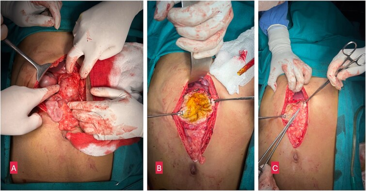 Figure 1
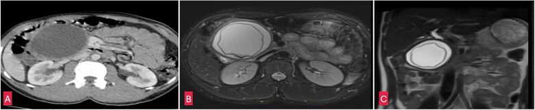 Figure 2
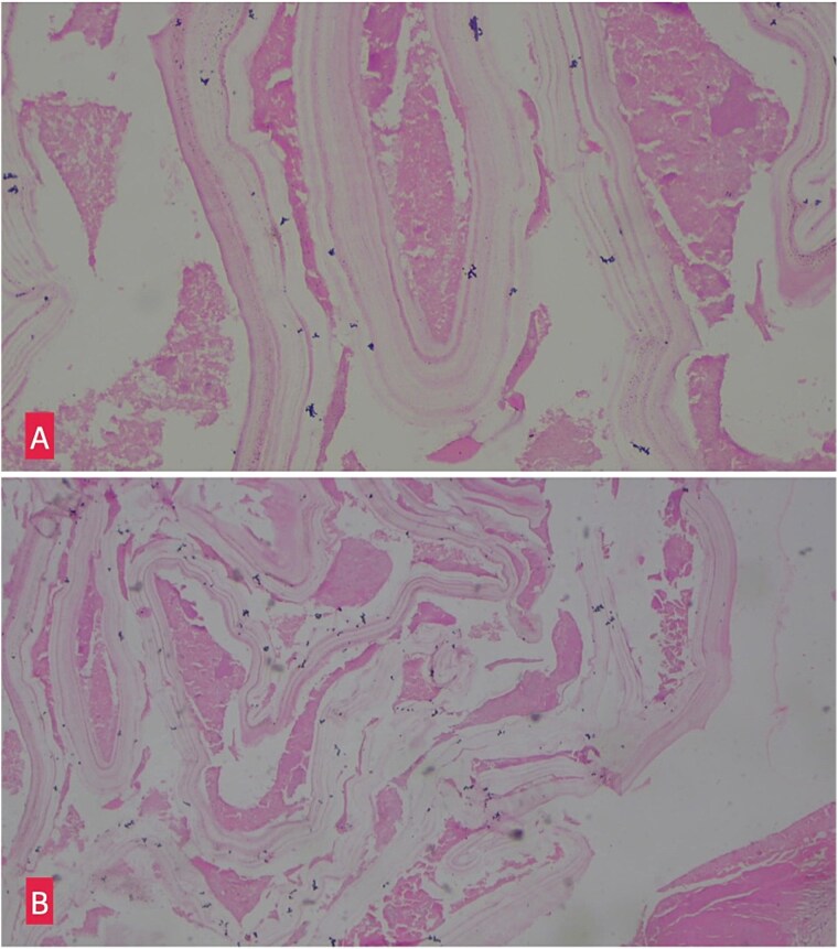 Figure 3
Peer Reviews
No public reviews on file for this paper yet. If you reviewed it on a platform where reviews are public (OpenReview, ICLR, NeurIPS, ICML), you can paste yours below so the community can read it here.
Videos
No videos yet. Explain this paper in a talk, walkthrough, or lecture? Add one.
Taxonomy
TopicsParasitic infections in humans and animals · Congenital Anomalies and Fetal Surgery · Genetic and Kidney Cyst Diseases
Introduction
Hydatid disease is a parasitic infection which is prevalent primarily in areas where livestock farming and agriculture are widespread [1]. Four species of Echinococcus are responsible for hydatid disease in humans [2]. Of these, Echinococcus granulosus is the most frequently encountered. In its lifecycle, dogs are the definitive hosts, with sheep and goats serving as intermediate host. Humans are accidental, dead end host.
The liver is the primary site of infection followed by lung. Pancreatic involvement is extremely rare. It accounts for <2% of systemic cases with only 33 cases documented in literature since 2011 [3]. Among the pancreas, the head is most commonly affected, followed by the body and tail [4].
In this case, we present a rare instance of a primary pancreatic hydatid cyst (PHC) that was misidentified as a pancreatic pseudocyst and subsequent management.
Case report
A 23 year old male presented with a 6-month history of epigastric pain and nausea. The pain was gradual, continuous, non-progressive, and mild in intensity. The pain radiated to the back and was dull aching in character with no aggravating or relieving factors. There were no other significant symptoms. On physical examination, a firm 7 × 7 cm lump was palpated in the epigastric region and left hypochondrium. It was spherical with ill-defined margins, smooth surface and did not move with respiration. The mass was not contiguous with liver dullness. When the patient was positioned in the knee-elbow position the lump did not fall forward suggesting retroperitoneal location.
Blood tests were unremarkable and serum amylase (103 U/L) and lipase (165 U/L) were within normal limits ruling out acute pancreatitis. An abdominal ultrasound identified a cystic lesion in the head of the pancreas measuring 7 × 8 cm. Contrast enhanced computed tomography (CECT) whole abdomen revealed a unilocular, thick walled, hypodense cyst (7.5 × 8.2 × 10 cm) near the head and uncinate process of pancreas. The cyst displaced the antrum of the stomach, transverse colon, first and second part of the duodenum, suggestive of a pancreatic pseudocyst. However, the absence of trauma or pancreatitis made this diagnosis unlikely. MRI whole abdomen revealed a well defined, thick walled cyst with a characteristic undulating membrane suggesting hydatid cyst (Fig. 2). Echinococcal serology was positive, with IgG (16 U/ml) and IgM (20 U/ml) confirming the diagnosis of hydatid cyst.
The patient was diagnosed with a primary isolated PHC and underwent open partial pericystectomy and omentoplasty, along with perioperative albendazole therapy (15 mg/kg/day). During surgery, a large, thick-walled cyst (7 × 8 cm) was found in the head and uncinate process of the pancreas, displacing the duodenum and bulging through the transverse mesocolon (Fig. 1). The cyst was isolated using 10% povidone-iodine-soaked gauze, and decompression was performed by needle aspiration. A scolicidal agent (10% povidone-iodine) was used to sterilize the cavity before partial cystectomy (deroofing) was performed.
(A) Intraoperative image showing a large cystic lesion in the head of pancreas. (B) Opened up cyst after partial pericystectomy. (C) Omentoplasty.
(A) Coronal section of CECT abdomen showing a thick walled, well defined round to oval cystic lesion ~7.5 × 8 × 10 cm in the head and uncinate process of pancreas. (B) Coronal section of MRI-magnetic retrograde cholangio-pancreatography (MRCP) abdomen showing a well defined thick walled cystic lesion in the head and uncinate process of the pancreas with undulating membrane noted within the cyst. (C) Saggital section of MRI-MRCP abdomen with similar findings as above.
(A) Hemotoxylin and eosin ×10: Section shows lamellated appearance of hydatid cyst. (B) Hemotoxylin and eosin ×40: Section shows lamellated appearance of hydatid cyst.
Histopathological examination of the excised cyst wall confirmed the diagnosis, showing a lamellated membrane (Fig. 3).
Postoperative period was uneventful and the patient was discharged on oral albendazole therapy for 8 weeks. He had made a full recovery and is doing well on follow-up visits.
Discussion
Humans do not play a direct biological role in the life cycle of E. granulosus and are accidentally infected by ingesting eggs found in the feces of infected dogs. Upon ingestion, the eggs pass through the intestinal wall, enter the portal circulation, and ultimately lodge in liver [5]. The most common mechanism of spread being hematogenous dissemination [6]. Other pathways include through the biliary system, lymphatic spread, or direct migration [6].
The growth of these cysts is slow, averaging 0.3–2 cm per year [7], and many patients remain asymptomatic for years.
Hydatid cysts in the head of the pancreas may present with obstructive jaundice or acute pancreatitis. Cysts in the body of the pancreas are usually silent until they grow large enough to cause abdominal distension, nausea, vomiting, and abdominal pain. On rarer occasions, cysts located in the tail may lead to splenomegaly or portal hypertension [7].
Ultrasonography is a non-invasive and cost-effective tool that is often the first choice, but its utility is limited in pancreatic cysts due to the retroperitoneal location of the pancreas and the interference from bowel gas. CT scan is more effective for determining cyst size, location, and its relationship to the pancreatic and biliary systems. MRI is particularly useful for evaluating the cyst's relationship with the bile ducts and pancreas [8].
Surgical intervention remains the primary treatment for PHCs, and the approach depends on the cyst's location.
Conclusion
Primary pancreatic hydatid cyst is a rare entity that can masquerade as pseudocyst or cystic neoplasm making the diagnosis challenging. The prognosis is usually favourable when appropriately managed. Surgical intervention combined with albendazole remains the main stay of treatment.
The reference list from the paper itself. Each links out to its DOI / PubMed record.
- 1Ahmed Z, Chhabra S, Massey A, et al. Primary hydatid cyst of pancreas: case report and review of literature. Int J Surg Case Rep 2016;27:74–7. 10.1016/j.ijscr.2016.07.054.27552034 PMC 4995534 · doi ↗ · pubmed ↗
- 2Akbulut S, Yavuz R, Sogutcu N, et al. Hydatid cyst of the pancreas: report of an undiagnosed case of pancreatic hydatid cyst and brief literature review. World J Gastrointest Surg 2014;6:190–200. 10.4240/wjgs.v 6.i 10.190.25346801 PMC 4208043 · doi ↗ · pubmed ↗
- 3Kothiya PK, Gupta V, Sarawagi R, et al. Isolated primary hydatid cyst of the pancreas: management challenges of a cystic masquerade. Ann Hepatobiliary Pancreat Surg 2022;26:401–6. 10.14701/ahbps.22-031.35995585 PMC 9721249 · doi ↗ · pubmed ↗
- 4Wani RA, Wani I, Malik AA, et al. Hydatid disease at unusual sites. Int J Case Reposts Images 2012;3:1–6. 10.5348/ijcri-2012-06-128-RA-1. · doi ↗
- 5Shah OJ . Hydatid cyst of the pancreas. An experience with six cases. JOP 2010;11:575–81.21068489 · pubmed ↗
- 6Mandelia A, Wahal A, Solanki S, et al. Pancreatic hydatid cyst masquerading as a choledochal cyst. J Pediatr Surg 2012;47:e 41–4. 10.1016/j.jpedsurg.2012.07.054.23164030 · doi ↗ · pubmed ↗
- 7Eris C, Akbulut S, Yildiz MK, et al. Surgical approach to splenic hydatid cyst: single center experience. Int Surg 2013;98:346–53. 10.9738/INTSURG-D-13-00138.1.24229022 PMC 3829062 · doi ↗ · pubmed ↗
- 8Makni A, Jouini M, Kacem M, et al. Acute pancreatitis due to pancreatic hydatid cyst: a case report and review of the literature. World J Emerg Surg 2012;7:7. 10.1186/1749-7922-7-7.22445170 PMC 3325852 · doi ↗ · pubmed ↗