Therapeutic Whole Lung Lavage for Pulmonary Alveolar Proteinosis: Technique With Continuous Video‐Enabled Double Lumen Endotracheal Tube
Kevin Davidson

TL;DR
A new technique using a special endotracheal tube makes lung lavage safer for a rare lung disease called autoimmune pulmonary alveolar proteinosis.
Contribution
Introduces a safer method for therapeutic whole lung lavage using a continuous video-enabled double-lumen endotracheal tube.
Findings
The technique improves patient safety during therapeutic whole lung lavage.
It is effective for treating autoimmune pulmonary alveolar proteinosis.
Abstract
Therapeutic whole lung lavage with a continuous video‐enabled double‐lumen endotracheal tube improves the safety of therapeutic whole lung lavage in autoimmune pulmonary alveolar proteinosis. Autoimmune pulmonary alveolar proteinosis is a rare lung disease which causes dyspnoea, cough, and subsequent respiratory failure due to the accumulation of surfactant proteins in the lung. Using a continuous video‐enabled double‐lumen endotracheal tube, we demonstrate how this procedure can be done with significantly improved patient safety.
Genes, proteins, chemicals, diseases, species, mutations and cell lines named across the full text — each resolved to its canonical identifier and authoritative record.
Click any figure to enlarge with its caption.
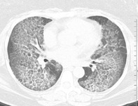 Figure 1
Peer Reviews
No public reviews on file for this paper yet. If you reviewed it on a platform where reviews are public (OpenReview, ICLR, NeurIPS, ICML), you can paste yours below so the community can read it here.
Videos
No videos yet. Explain this paper in a talk, walkthrough, or lecture? Add one.
Taxonomy
TopicsNeonatal Respiratory Health Research · Congenital Diaphragmatic Hernia Studies · Neuroscience of respiration and sleep
Case Video
1
Autoimmune pulmonary alveolar proteinosis is a rare lung disease which causes dyspnoea, cough, and subsequent respiratory failure due to the accumulation of surfactant proteins in the lung [1]. Isolating each lung sequentially under anaesthesia to perform therapeutic whole lung lavage is a proven technique to treat this condition [1, 2]. However, there are substantial risks of performing whole lung lavage including double‐lumen endotracheal tube migration, challenges with isolating each lung for high‐volume irrigation or ventilation, and hypoxemia [1, 2]. Using a continuous video‐enabled double‐lumen endotracheal tube, we demonstrate how this procedure can be done with significantly improved patient safety (Video 1).
The video highlights the key images in proper double‐lumen endotracheal tube placement during sequential therapeutic whole lung lavage of the left and right lungs. Viewers are shown key technical aspects of this procedure, which is performed for a rare lung disease: pulmonary alveolar proteinosis. The procedure can be life‐saving and is infrequently performed. Patient safety is improved by continuous video monitoring of tube placement throughout the entire procedure to reduce the risk of complications. Video content can be viewed at https://onlinelibrary.wiley.com/doi/10.1002/rcr2.70153
Author Contributions
Kevin Davidson owns this video, wrote captions and the manuscript, performed edits and entered the submission.
Ethics Statement
The author declare that appropriate written informed consent was obtained for the publication of this manuscript and accompanying images.
Conflicts of Interest
The author declares no conflicts of interest.
The reference list from the paper itself. Each links out to its DOI / PubMed record.
- 1C. Mc Carthy , B. C. Carey , and B. C. Trapnell , “Autoimmune Pulmonary Alveolar Proteinosis,” American Journal of Respiratory and Critical Care Medicine 205, no. 9 (2002): 1016–1035.10.1164/rccm.202112-2742 SOPMC 985147335227171 · doi ↗ · pubmed ↗
- 2A. Awab , M. S. Khan , and H. A. Youness , “Whole Lung Lavage‐Technical Details, Challenges, and Management of Complications,” Journal of Thoracic Disease 9, no. 6 (2017): 1697–1706.28740686 10.21037/jtd.2017.04.10PMC 5506114 · doi ↗ · pubmed ↗