Correction: l-Arginine, as an essential amino acid, is a potential substitute for treating COPD via regulation of ROS/NLRP3/NF-κB signaling pathway
Chunhua Ma, Kexi Liao, Jing Wang, Tao Li, Liangming Liu

Abstract
Genes, proteins, chemicals, diseases, species, mutations and cell lines named across the full text — each resolved to its canonical identifier and authoritative record.
Click any figure to enlarge with its caption.
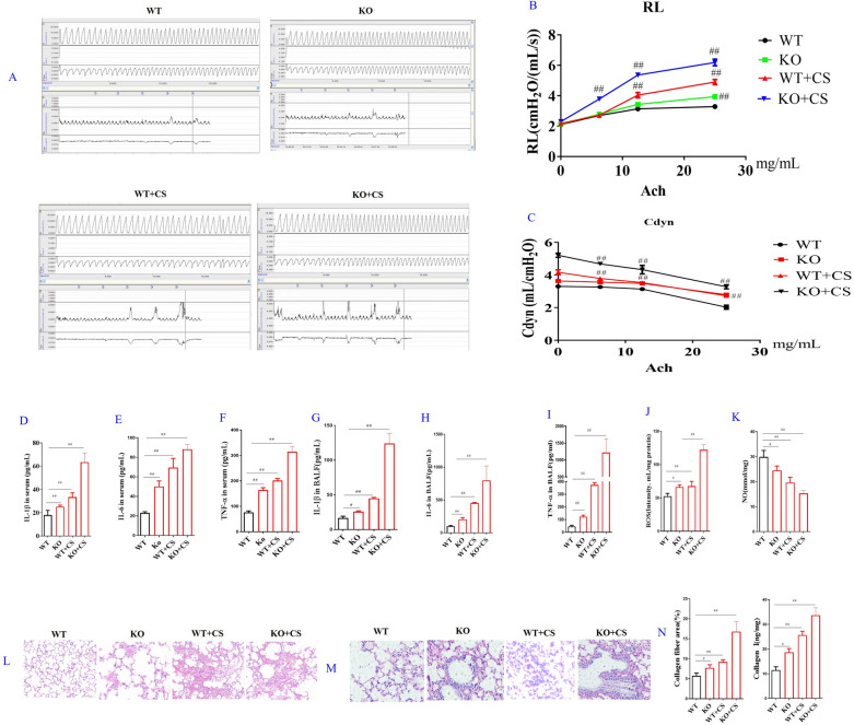 Figure 3
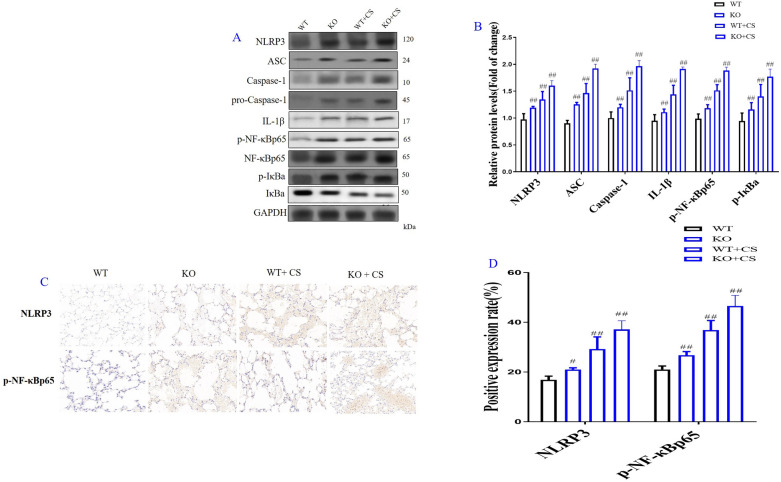 Figure 7
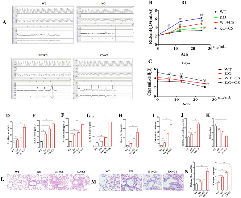 Figure 3
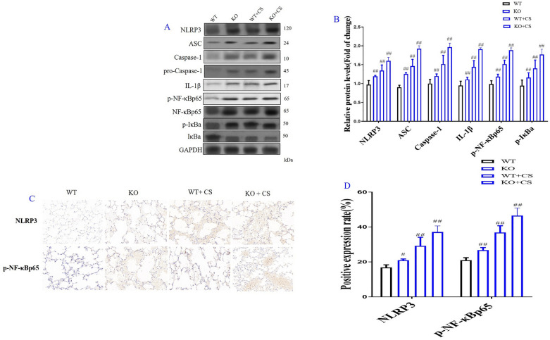 Figure 4
Peer Reviews
No public reviews on file for this paper yet. If you reviewed it on a platform where reviews are public (OpenReview, ICLR, NeurIPS, ICML), you can paste yours below so the community can read it here.
Videos
No videos yet. Explain this paper in a talk, walkthrough, or lecture? Add one.
Taxonomy
TopicsChronic Obstructive Pulmonary Disease (COPD) Research · Pediatric health and respiratory diseases · Respiratory Support and Mechanisms
Correction: Cell & Bioscience (2023) 13:152 10.1186/s13578-023-00994-9
In this article [1], the wrong figures appeared as Figs. 3 and 7; the figures should have appeared as shown below.
Incorrect Fig. 3
Correct Fig. 3Fig. 3. Effects of l-arginine (LA) on KO COPD mice. Airway reaction (A). The percentage changes of the resistance of lung (RL) (B) and lung dynamic compliance (Cdyn) (C) in WT and KO COPD mice. Serum cytokines: The contents of interleukin-1β (IL-1β) (D), interleukin-6 (IL-6) (E), tumor necrosis factor-α (TNF-α) (F). BALF cytokines: the contents of Interleukin-1β (IL-1β) (G), interleukin-6 (IL-6) (H), tumor necrosis factor-α (TNF-α) (I). Reactive oxygen species (ROS) (J) and nitric oxide (NO) (K) contents in lung tissues. Pathological changes (HE staining) and Masson staining of lung in COPD rats: HE staining of lung in COPD rats (× 200) (L), Masson staining of lung in COPD rats (× 200) (M), Collagen quantification of Masson staining and collagen I contents of lung in COPD KO mice (N). (n = 10). All data were presented as mean ± SD. Compared with WT mice: ^##^P < 0.01
Incorrect Fig. 7
Correct Fig. 7Fig. 7. Role of l-arginine (LA) mediated ROS/NLRP3/NF-κB signaling pathway in cigarette smoke extract (CSE)-induced primary bronchial epithelial cell (BEC) injury and molecular docking of LA and NLRP3. Western blot of ROS/NLRP3/NF-κB signaling pathway in CES-induced BECs (A), Quantification of ROS/NLRP3/NF-κB signaling pathway in CES-induced BECs (B). The expression levels of NLRP3 (C) and p-NF-κBp65 (D) in CES-induced BECs by immunofluorescence (× 100). (n = 3). Molecular docking result of LA and NLRP3 (E): The binding energy predicted by Autodock is − 5.79 kcal/mol for LA-NLRP3 (The binding energy predicted by Autodock < − 6.00 is considered to be high degree of integration). All data were presented as mean ± SD. Compared with control: ^##^P < 0.01